The skull of a human being is an amazing piece of work in biology. It does not only offer a hard protective covering to the brain but also it is where the face structure is formed, a place where senses are housed and help in crucial functions like eating, breathing, and talking. Knowledge of the bones of the skull and face is the cornerstone of anatomy as well as medicine and practice. This paper examines their classification, names, locations, functions, and clinical relevance making the complex osteological concepts simpler to understand by students, healthcare providers, and any other person who would be interested in the anatomy of the human body.
Overview of Skull Anatomy
The skull is made up of facial bones and cranial bones, which unite to create a structure that can safeguard the brain besides supporting the face. There are 22 skull bones in an adult human being, without the middle ear ossicles. Sutures hold these bones together, non-portable joints which gradually become fused during the course of age.
Functions of the Skull
The skull plays the major roles of:
- Protection: The brain is enclosed in a hard protective case that is made up of the cranial bones.
- Support: The bones of the face form a support of the soft tissues, muscles, and organs.
- Housing of Sensory Organs: The sockets of the eyes, nose and ear structures are covered by the skull.
- Communication: The speech and mastication are facilitated by the jaw and the mouth.
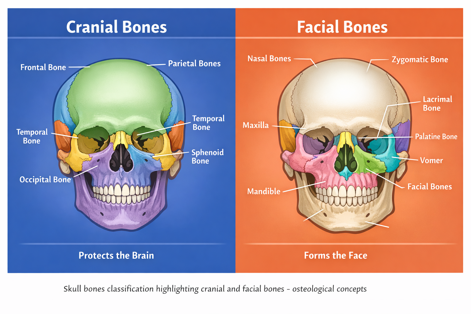
The Cranial Bones are classified into four groups
The skull consists of bones that create the protection of the brain. These are separated into paired and unpaired bones, and there are eight of them.
Unpaired Cranial Bones
Frontal Bone
- Location: Develops the forehead and the top of the eye sockets (orbits).
- Function: Covers the frontal lobes of the brain and undergoes facial muscles.
- Clinical significance: Fractures may cause cosmetic anomalies or brain damage.
Occipital Bone
- The point of location: back of the skull.
- Role: Covers the cerebellum and the brainstem; creates the foramen magnum of the spinal cord.
- Repeatedness: Trauma can result in the compression of the brain stem, or impairment of the cranial nerves.
Sphenoid Bone
- Location: The central skull base with an interconnection with practically all other bones of the head.
- Function: This offers the gland of structural stability and it contains the pituitary gland in the silla turcica.
- Clinical significance: Fracture of the sphenoid may affect the vision and hormonal processes.
Ethmoid Bone
- Location: This is located in-between the orbits and a component of the nasal cavity and anterior cranial fossa.
- Use: The nasal cavity is supported and the medial walls of the orbit contributed by it.
- Clinical implications: Fractures can cause a leakage of cerebrospinal fluid or orbital traumas.
Paired Cranial Bones
Parietal Bones (2)
- Location: almost all of the skull, including the sides and the top.
- Purpose:Guard the parietal lobes of the brain.
- Clinical significance: Prone to demoralizing trauma resulting in epidural hemorrhages.
Temporal Bones (2)
- Location: Areas of the skull towards the side of ears.
- Function: The inner ear and the middle ear contain houses that support the muscles of mastication.
- Clinical implications: Hearing, balance, and facial nerve impairment may be the outcomes of fractures.
Facial Bones classification
The anterior structure of the skull is made by facial bones who support eyes, nose and mouth. The number of bones that make the face is 14 with six being matched and two not. Facial bones have been categorized into the following:
Unpaired Facial Bones
Mandible
- Location: Lower jaw.
- Role: helps to support lower teeth, masticate and speak.
- Clinical significance: In trauma, this fracture is frequently broken; the inappropriate healing may hamper the chewing and occlusion.
Vomer
- Location: The location constitutes the back of the nasal septum.
- Function: Partition of the nasal cavity into two nostrils.
- Clinical impact: The deviation can lead to problems with breathing and be a cause of sleep apnea.
Paired Facial Bones
Maxillae (2)
- Location: Upper jaw and mid facial.
- Function: To support the upper teeth, shape the part of the orbit, and the nasal cavity as well as the hard palate.
- Clinical relevance: The fractures of the maxilla can impact the vision, occlusion, and nasal functioning.
Zygomatic Bones (2)
- Location: Cheekbones.
- Function: The cheeks are given prominence, and they make a part of the orbit.
- Clinical relevance: Fractures may result in finding of the face and eye problems.
Nasal Bones (2)
- Location: Bridge of the nose.
- Function: nose cartilage of the support.
- Clinical significance: It is commonly broken in facial injuries; it can be cosmetically deformed.
Lacrimal Bones (2)
- Position: Within the orbits of the middle of the walls.
- Role: safeguard lacrimal sacs which helps the drainage of tears.
- Clinical relevance: Teardrop injuries can result in hindrance by the tearflow or chronic tearing.
Palatine Bones (2)
- Location: The second one is located in the back of the palate and the floor of the nasal cavity.
- Function: This is a component of the oral and nasal cavities.
- Clinical implications: The cleft palate or speech problems may be brought up by malformation.
Inferior Nasal Conchae (2)
- Location Lateral nasal cavity walls.
- Function: To increase the area of humidification and filtration of the air which was inhaled.
- Clinical significance: Structural problems may play the role of acute nasal obstruction.
Anatomical Significance of Skull and Facial Bones
Fractures
- Cranial fractures: In most cases, they are caused by blunt trauma; can be followed by intracranial hemorrhage, brain contusion, or injury of cranial nerves.
- Facial fractures: They include Le Fort maxilla fractures, zygomatic fractures, and nasal bone fracture. These injuries may affect the vision, breathing, mastication and aesthetics.
Congenital Deformities
- Craniosynostosis: This is the premature closing of the cranial sutures and it also can have the effect on the shape of the skull and possibly the brain development.
- Cleft lip and palate: This is due to the partial fusion of facial bones during development and it affects feeding, speech and dentition.
Neurological and Facial Functional Effect
- Cranial bone trauma or deformities may have a direct impact on the functioning of the brain, such as cognition, motor control, or sensory processing.
- The disorders of the facial bones could affect asthma, eyesight, or chewing. As a case in point, zygomatic fractures may decrease the orbital volume, which in turn may influence the eye movements and vision.
Application of Imaging in Clinical Practice
Skull and facial bone integrity assessment can only be done using modern imaging methods like CT scans and MRI. They assist in identification of fractures, evaluation of deformities and surgical planning. Imaging in trauma cases is used to make sure that neurological infringement or irreparable facial dysfunction is prevented in time.
The Essentials of Osteology
Skull and facial bones are studied incorporating a number of osteological notions:
- Classification of bones (paired/unpaired, flat/irregular)
- Suture morphology (coronal, sagittal, lambdoid)
- Adaptation of functions (thickened areas where muscles are attached, sinus cavities in order to lose weight)
The knowledge of such concepts enables healthcare providers to predict the occurrence of clinical complications and design effective interventions. Visualization of bones in three dimensions can help students of anatomy to understand the protective and functional roles of the bones.
Conclusion
The skull of a human being is a masterpiece of evolutionary designs with its ability to be strong, offer security, and be pleasing to the face. The cranial bones protect the brain, whereas the facial bones make the face and help to perform such important functions as seeing, grinding, and breathing. The familiarity with these bones and their clinical significance is essential to the knowledge of the trauma management, postnatal and congenital anomalies, and surgical operations.
Reducing the complexity of osteological concepts, health care workers, students, and amateurs will be able to enjoy the complexity of connections between bone structure, functioning, and the clinical outcomes. The acknowledgement of the way in which fractures, deformities, or developmental conditions can influence either neurological or facial functions is a guarantee of good diagnosis and subsequent treatment.