Oral Radiology and its importance in dentistry.
Oral Radiology is a specialized area of dentistry used to diagnose, monitor, and direct treatment for all conditions involving the teeth, jaws and associated oral structures utilizing imaging systems. It is very important for modern teeth health as many tooth diseases and abnormalities will remain undetected by just looking at them. These are different types of radiographs that will enable your dentist to look under the surface gums and teeth, helping to detect problems early on, including any tooth decay, bone loss, infection, and/or developmental abnormalities. This means that it is a valuable asset to the fields of preventive dentistry, restorative planning and long-term oral health management.
Advancements in digital imaging and 3D scanning along with computer-assisted diagnostics are constantly bringing new developments to the field, transforming dental care into more precise and efficient processes than ever before. Knowledge of oral radiology offers insights to students, patients, and practitioners about the groundbreaking nature of modern dentistry, which has come to rely on precise diagnosis and successful treatment. Advancements in dental technologies have made imaging a key component of the clinical decision making process that ensures that the practice can provide safer, more predictable and more individualized care for patients of all ages.
Oral radiology: What is it? A Foundational Overview
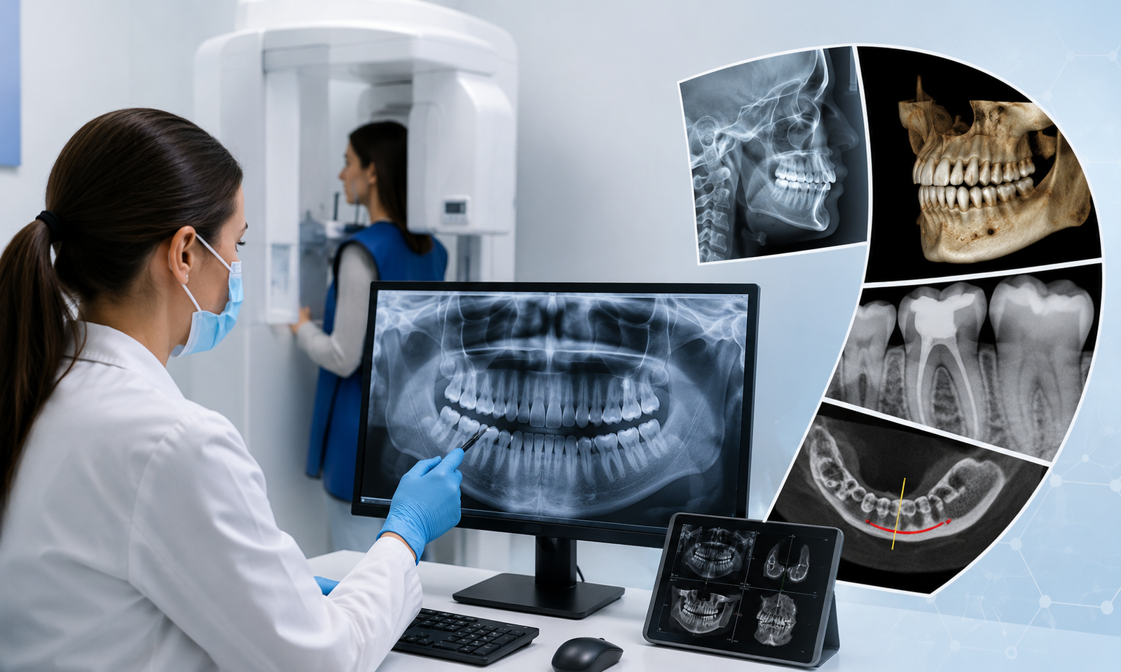
Oral radiology is the TREATMENT and science of X-rays and other imaging techniques to look after the structures of the oral cavity and the face. It consists of methods including periapical radiographs, bitewing X-rays, panoramic imaging and advanced 3D cone-beam computed tomography (CBCT). Thanks to these tools, dentists can better see what’s going on inside the oral cavity, such as with the roots of the teeth, jawbone density, impacted teeth and underneath infections. To appreciate the role of oral radiology in connecting with clinical oral medicine and science in areas of diagnosis and making informed decisions.
Oral Radiology is not just about taking pictures. It includes reading of X-rays, recognition of abnormalities and linking radiological data to symptoms. Healthcare professionals involved in this area of training will be able to identify the small changes which might occur at the start of disease. Oral radiology has become a necessity in dental education and practice. The study of radiographic interpretation is crucial, especially for incoming dental students, since it affects diagnosis and treatment, which in turn has a direct influence on achieving a successful treatment outcome.
Dental Imaging in Modern Dental Health Care
The introduction of dental imaging has revolutionized the field of dentistry and provided a crucial tool that has improved the accuracy of diagnosis and the efficiency of treatment. Many dental conditions would not be discovered because radiographs were not taken. For instance, a simple tooth decay may occur between teeth and under existing fillings that are concealed in a routine checkup. Dentists can better understand such issues with the use of radiographic imaging, avoiding the future need of extensive treatment and complications.
Dental imaging is also vital in the diagnosis of periodontal disease, root infection, cysts, tumours and injuries created by trauma to the teeth. It can also be used as a good indicator of tooth alignment, and for more insight into the development of permanent dentition in children and adult growth. Imaging provides a detailed look inside the mouth and a basis for making conservative, effective treatment decisions for the maintenance side of dentistry. This helps to increase patient satisfaction which in turn diminishes oral health risks in the future.
Types of radiographic techniques used in dentistry
In the field of oral radiology, there are a number of radiographic procedures, and each has a particular diagnostic application. Intraoral radiographs (periapical and bite wings) frequently are used for cavity detection, bone level evaluation and individual tooth assessment. Bitewing X-rays can effectively reveal decay between the teeth, and periapical X-rays can give a detailed look at the full tooth structure, such as the root and supporting bone.
Extraoral imaging techniques like panoramic radiographs give an overall picture of the whole mouth, both jaws, sinus and the temporomandibular joints. It is particularly beneficial in evaluating misaligned teeth, abnormal development and impacted teeth. Advanced imaging techniques, including cone-beam computed tomography (CBCT), provide a three-dimensional view and enable precise diagnosis and analysis of complex scenarios, such as dental implant planning, orthodontic treatment and surgical procedures. All these techniques make their contribution to diagnosis in a unique manner, which helps dentists to have as much knowledge as possible about the condition.
Role of Oral radiology in Diagnosis of disease.
Dental and maxillofacial diseases are linked to oral radiology with one of the most crucial roles being the early detection and diagnosis of dental and maxillofacial disease. The majority of conditions start without any significant signs and symptoms, this is why early intervention with a radiographic examination is crucial. In the case of dental caries, for example, there could be no obvious pain, but may be under a part of the tooth’s enamel surface or between teeth. Early restorative treatment can be performed on these hidden lesions in order to prevent the injury from becoming widespread with the help of a radiograph.
The role of oral radiology is important especially for the diagnosis of bone infections (osteomyelitis), cystic lesions, benign or malignant tumors. Sometimes more extensive imaging might be necessary to find the size, position, and relationship to other structures or tissues through which these conditions may evolve. Radiographs give the dentist a good idea of what is going on internally within the oral cavity, helping him or her determine the type of pathology and determine whether additional imaging tests or referral to a specialist is indicated. This greatly enhances the safety of patients and treatment outcomes.
Treatment planning and clinical decision making.
A set of all the procedures that are performed in the treatment planning process is known as a road map.In dentistry, a road map is a collection of all the procedures to be used in treatment planning. Radiographic evaluation allows for the determination of the most suitable treatment for a patient, whether they need fillings, root canal treatment, orthodontics or dental implants. In restorative dentistry, for instance, radiographical information will inform the dentist of the degree of decay along with how much tooth structure can be retained as part of the treatment.
The imaging may also be necessary when oral surgery or implant placement is planned to properly assess the bone density, position the nerves, and understand any limitations. This allows the procedures to be done in a safety oriented manner, with minimal risks of complications. Radiographic analysis is also an important tool for orthodontists in determining the relationships of the jaws and tooth alignment prior to treatment plan design. In summary, oral radiology is a vital tool that enables dentists to tailor treatment to each patient’s unique needs and objectives, optimizing the likelihood of successful results.
Identifying Oral and Dental issues
Various dental and maxillofacial disorders tend to be hidden and not evident in the visual examination. Common problems that oral radiology helps to identify include impacted teeth, fracture of the roots, infection of the gums (abscesses) and bone defects. Supporting illustrations of impacted wisdom teeth are common, for example, when wisdom teeth are found by panoramic x-rays before they cause pain or infection.
In addition, it aids in uncovering alterations in the bone’s structure, which may be associated with systemic disease or chronic infection. Sometimes low bone density seen on the radiograph of the jaws, which could be associated with osteoporosis, can help identify the condition. Such problems can be diagnosed when they are early and initially treated to prevent any further complications from arising. This diagnostic secret is one of the greatest assets that oral radiology has provided to the care of the public.
Structures of the jaws and facial development.
Patients with dental pain may be further assessed with oral radiology, which goes hand in hand with the complex anatomy of the jaw and surrounding facial structures. Mandibles and maxillae are essential for chewing, speech and facial appearance which are vital in dental care. On the other hand, radiographic imaging enables the doctor to assess bone quality, rule out fractures, and assess the condition of the temporomandibular joints (TMJ).
Detailed images are used in maxillofacial surgery and orthodontics to evaluate the alignment and symmetry of the face and jaws. This information becomes indispensable in the design of corrective procedure and/or reconstructive surgery. Oral radiology enables practitioners to plan the treatment of dental disease accurately and effectively, due to the fact that it gives them a complete picture of the teeth’s skeletal relationships. It also aids in the recovery of bone and tissue following surgery and lets surgeons know how well things are going.
Patient Care, Safety and Radiation Considerations
Radiography used in dentistry on the contrary exposes the patients to relatively low amounts of radiation but the patient’s safety is still given utmost care and protected through the systematic principles and protocols while still practicing radiology in dentistry. Digital imaging systems have lowered the amount of radiation needed, compared with the use of film. Plus, lead aprons, thyroid collars, and other protective measures are routinely implemented to reduce exposure.
Dentists use the principle of ALARA to put as few radiographs as reasonably possible in the mouth.Dentists strive to adhere to the As Low As Reasonably Achievable (ALARA) principle when deciding to take radiographs, making sure that the radiographs are only taken when medically necessary. Careful approach that weighs benefits of diagnosis against the safety of the patient. New technology is constantly emerging that makes dental imaging more efficient and safer, with a reduced dose of radiation and better quality pictures. Education of patients is also a critical part of oral radiology, enabling people to comprehend the need and safety of diagnostic and therapeutic radiography.
Future of oral radiology in Dentistry
Oral radiology’s future is intertwined with the advancements in digital technology, artificial intelligence and 3D imaging systems. The field of dentistry is witnessing a new wave of AI-driven diagnostic tools designed to facilitate a more precise and efficient interpretation of radiographs for dentists.The diagnostic field is seeing a new era of AI-powered products that would enable dentists to interpret radiographs more accurately and efficiently. These systems can point out possible irregularities, minimize misdiagnosis, and enhance medical choices.
In addition, 3D imaging and virtual planning are revolutionizing more complicated treatments including those of implant placement and orthodontic procedures. In the future, oral radiology will increasingly become a part of digital dentistry, making it easier to manage with greater accuracy and detail to enhance patient care. In conclusion, for both students and professionals, it is essential to understand these changes and be ready for dental care in the future.


