Modern implant dentistry has evolved significantly with the introduction of advanced imaging technologies that enhance accuracy, efficiency, and patient comfort. These tools allow clinicians to visualize anatomical structures with remarkable clarity, plan treatments with greater precision, and collaborate more effectively with laboratories. As digital workflows continue to expand, imaging technologies have become essential components of successful implant planning and placement. Understanding the innovations shaping this field highlights how dentistry is moving toward more predictable and patient centered care.
Cone Beam Computed Tomography for Three Dimensional Clarity
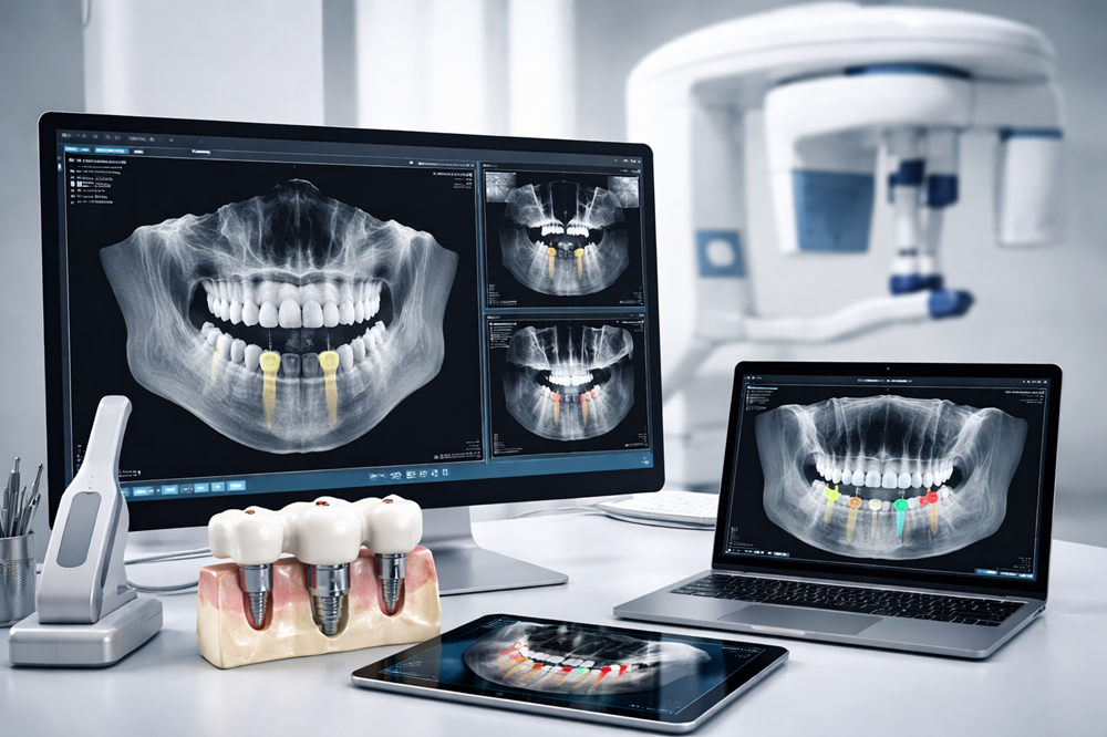
Cone beam computed tomography has become one of the most important imaging tools in implant dentistry. Unlike traditional two dimensional radiographs, CBCT provides a detailed three dimensional view of the patient’s bone structure, nerve pathways, and surrounding tissues. This level of detail allows clinicians to evaluate implant sites with exceptional accuracy and identify potential challenges before treatment begins.
CBCT imaging supports safer and more predictable outcomes by helping providers assess bone density, determine ideal implant positioning, and visualize anatomical variations. The ability to rotate and examine the scan from multiple angles enhances diagnostic confidence and strengthens the foundation for treatment planning.
Intraoral Scanners for Digital Impression Accuracy
Intraoral scanners have transformed the way impressions are captured in modern dentistry. These devices create highly accurate digital models of the teeth and soft tissues without the need for traditional impression materials. Patients benefit from a more comfortable experience, while clinicians gain access to precise data that integrates seamlessly with digital workflows.
Digital impressions reduce the risk of distortion and allow for faster communication with laboratories. The resulting models support improved restorative design, better fitting prosthetics, and a more efficient overall process. Intraoral scanning has become a cornerstone of digital implant planning due to its reliability and ease of use.
Panoramic Imaging for Broad Diagnostic Insight
Panoramic imaging remains a valuable tool in implant dentistry, offering a comprehensive view of the upper and lower jaws in a single image. While not as detailed as CBCT, panoramic radiographs provide essential information about tooth positioning, bone height, and overall oral health.
This broad perspective helps clinicians identify potential concerns early in the planning process. Panoramic imaging is often used as an initial diagnostic tool before more advanced imaging is performed. Its ability to capture a wide field of view makes it a practical and efficient part of modern implant workflows.
Photogrammetry for Full Arch Precision
Photogrammetry has become an increasingly important technology in full arch implant dentistry. This method captures the exact spatial relationship between implants using specialized cameras and software. The resulting digital models are highly accurate and support precise restorative design.
Practices that incorporate reputable full arch dental photogrammetry benefit from reduced chair time, improved fit of prosthetics, and a more streamlined workflow. This technology is especially valuable in complex cases where accuracy is essential for long-term success. By eliminating the need for traditional impressions in full arch restorations, photogrammetry enhances both efficiency and patient comfort.
3D Printing for Custom Surgical Guides and Models
Three dimensional printing has expanded the possibilities for personalized implant care. Using digital data from CBCT scans and intraoral impressions, clinicians can create custom surgical guides that support precise implant placement. These guides help ensure that implants are positioned at the correct angle, depth, and location based on the treatment plan.
Beyond surgical guides, 3D printing is used to fabricate models, provisional restorations, and other components that support communication and planning. This technology enhances accuracy and reduces turnaround times, making it a valuable addition to modern implant imaging workflows.
Digital Treatment Planning Software for Predictable Outcomes
Advanced treatment planning software brings together data from multiple imaging sources to create a comprehensive digital plan. Clinicians can simulate implant placement, evaluate bone quality, and visualize the final restoration before any surgical steps are taken. This level of planning reduces uncertainty and supports more predictable outcomes.
Digital planning software also enhances collaboration between clinicians and laboratories. When both teams work from the same detailed digital models, communication becomes more efficient and restorative results become more consistent. This integration strengthens the entire workflow from diagnosis to final placement.
Conclusion
Modern implant imaging relies on a combination of advanced technologies that enhance accuracy, efficiency, and patient comfort. CBCT, intraoral scanners, panoramic imaging, photogrammetry, 3D printing, and digital planning software each play a vital role in creating predictable and streamlined workflows. As these tools continue to evolve, they support a higher standard of care and contribute to long lasting, successful implant outcomes.