Spinal cord happens to be one of the most essential parts of the human nervous system. It is the primary communication pathway between the brain and the rest of the body in which sensory input and motor output travel up and down, respectively, to the brain and muscles/organs respectively. The spinal cord, outside of communication, is also a vital protective system, as it allows the reflexive response, or rapid, automatic reactions that can be used to avoid severe injury. Knowledge of the construction of the spinal cord is fundamental to the student of biology, anatomy, and health sciences and anybody who is interested in the mechanisms by which the body coordinates movement and sensation.
This article will discuss the structure of the spinal cord, its parts, the arrangement of the gray and white matter, the major nerve pathways, and how the spinal cord is involved in the action of the reflex.
Introduction to the Anatomy of the Spinal Cord
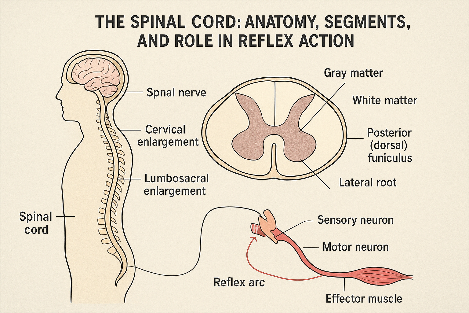
The spinal cord is a long, narrow tract of nervous tissue, which runs away out of the bottom of the brain (medulla oblongata) all the way down the back of the body, usually stopping somewhere between the first and tenth lumbar vertebrae in adults. It is located in the vertebral canal and it is covered by the vertebral column, meninges and the cerebrospinal fluid.
The fact that specific structure of the spinal cord are organized in a very clear manner can be used to understand how nerve impulses are effectively passed through and why the impairment of certain parts results in specific sensory or motor limitations.
Place and Protective Coatings
Location in the Spinal Column
The spinal cord is placed vertically in the vertebral canal that is created by a sequence of vertebrae. Although the column of the spine extends to the coccyx, the spinal cord stops at a point referred to as the conus medullaris. Under this, there are lower spinal nerves which are bundled together and this is known as the cauda equina.
Protective Layers
Meninges is a set of three layers of connective tissue which protects the spinal cord:
- Dura mater- tough outer layer.
- Arachnoid matter – the middle layer, a web-like one.
- Pia mater – the thin inner cover which is very closely attached to the cord.
Cerebrospinal fluid is found between arachnoid and pia mater, and provides cushioning to the spinal cord and minimizes the effects of sudden movements.
Segments of the Spinal Cord
The spinal cord is functionally separated into parts although they seem connected. Each part originates two spinal nerves one on the left and one on the right, that leave the vertebral canal via intervertebral foramina.
Major Spinal Cord Regions
The spinal cord is subdivided into 5 major sections depending on the spinal nerves that are emanated out of them:
Cervical region (C1-C8)
- Nourishes the neck, shoulders, arms and hands.
- The nerves in the upper limbs important in movement and sensation are found here.
Thoracic region (T1-T12)
- Supplies the abdominal muscles and the chest.
- The major role in the stability of the trunk and posture.
Lumbar region (L1-L5)
- Nourishes the lower back, hips and the legs.
- Significant in regard to walking and lower limb strength.
Sacral region (S1-S5)
- Manages sections of the pelvis, legs and feet.
- Manages bowel, bladder and sexual control.
Coccygeal region (Co1)
- Irrigates the peri-tailbone locality.
All the segments are associated with a certain dermatome (skin location) and myotome (muscle group), this is why spinal damages influence certain areas that can be predicted in the body.
The Spinal Cord, Inner Anatomy
An internal structure of the spinal cord is very organized that can be seen as a cross-section view designed to transmit signals rapidly.
Gray Matter Organization
The gray matter is seen in the middle of the spinal cord and is in the shape of a butterfly or H. The major components of the gray matter are neuron cell bodies, dendrites, and synapses.
The gray matter is parted into horns:
Posterior (dorsal) horns
- Perceive peripheral information in receptors.
- Process signals are associated with pain, temperature, and touch.
Anterior (ventral) horns
- Contain motor neurons
- Transmit messages to the skeletal muscles to get the skeletal muscles moving.
Lateral horns (are found in thoracic and upper lumbar areas)
- Included autonomic motor neurons.
- Control the automatic system like the heart rate and the digestion.
White Matter Distribution
The white matter surrounds the gray matter and it contains a majority of myelinated axons. These axons constitute ascending and descending tracts that are long distance carriers of information.
The white matter is broken into three columns also known as funiculi:
- Posterior (dorsal) columns – transmit information about the senses.
- Lateral columns – transport sensory as well as motor information.
- Anterior (ventral) columns – contain mostly motor signals.
The Spinal Cord Nerve Pathways
The spinal cord is a two way communication system and it employs specialized nerve pathways to convey the messages.
Ascending (Sensory) Pathways
Some of the tracts, known as ascending tracts, transport sensory signals in the body to the brain. Significant sensorimotor circuits are:
Spinothalamic tract
- Conducts pain, temperature and crude touch.
Dorsal column-medial lemniscus tract
- Feels small size, shakenness, and proprioception (awareness of body positioning)
These are the pathways in which the brain interprets the sensations and reacts accordingly.
Descending (Motor) Pathways
Downwards tracts pass motor instructions of the brain to muscles. Notably, significant motor pathways are:
Corticospinal tract
- Regulates voluntary movements, particularly, doing the hands and fingers very accurately.
Extrapyramidal tracts
- Control position, balance, and tone of muscle.
Injury to descending tracts usually leads to a weakness or paralysis below the area of injury.
Spinal Nerves and Transmission of Signals
All the spinal nerves are composed of two roots:
- Dorsal (posterior) root – is a conduit of sensory to the spinal cord.
- Ventral (anterior) root – is a motor outflow of the spinal cord.
The dorsal root has a swelling known as the dorsal root ganglion where neuron cells of the sense are located. After the entry of sensory information in the spinal cord it can be transmitted to the brain or can be utilized by itself in response to produce a reflex.
Spinal Cord Role in Reflex Action
The spinal cord is one of the most striking organs in that it is capable of creating reflexes, which are quick, automatic reactions unthoughtful.
What Is a Reflex?
Reflex can be described as an automatic reaction to a stimulus that aims at safeguarding the body. The response to reflexes is quicker than the voluntary response since the higher processing centers of the brain are bypassed.
Components of a Reflex Arc
The components in a typical reflex arc are 5 in number:
- Receptor – reacts to a stimulus (e.g. heat or pain)
- Sensory neuron – transmits the impulse to the spinal cord.
- Integration center- found in the gray matter of the spinal cord.
- Motor neuron – conducts the signal of response.
- Effector-muscle or gland which produces the response.
As an illustration, when one touches a hot surface, the sensory neurons send the pain to the spinal cord, which sends the motor command to withdraw the hand immediately.
Types of Reflexes
- Stretch reflexes – keep the muscles in place and on their feet (e.g., kneejerk reflex)
- Reflexes of withdrawal – withdraw the body after a painful stimulus.
- Autonomic reflexes – control internal body organs, e.g., blood pressure and digestion.
These reflexes show that the spinal cord can respond, without the intervention of the brain to provide quick protection.
Spinal Cord Anatomy Clinical Significance
Knowledge of spinal cord anatomy is necessary in the medical and clinical professions. The severity and nature of functional loss depends on the level and extent to which there is damage on the spinal cord.
- Complete injury- loss of sensation and movement below the level of injury.
- Incomplete injury- partial retention of sensory or motor activity.
Another useful diagnostic method is reflex testing, which assists the clinician in determining the integrity of the nerves and identifying neurological conditions.
Conclusion
The spinal cord is much more than mere connection between the body and the brain. Its divided structure, complexity of gray and white matter and specific nerve passages make it possible to control the movement, sensation, and reflexes with accuracy. The knowledge about the spinal cord structure can help readers to comprehend the way the signals of the senses and movements move effectively and the way reflexes can save the body in case of immediate threat. This understanding will be the basis of further research in the field of anatomy, physiology and clinical neuroscience as the spine cord plays a crucial role in the normal functioning of the human body.