Oral mucosa refers to a special type of epithelial tissues that line the mouth cavity and are important in helping to protect underlying structures, as well as, to aid oral functions and to support oral health. Knowledge of the microscopic structure and variations of oral mucosa is vital to the clinicians, dental surgeons, and researchers. It assists in the diagnosis of diseases, the organization of surgical operations, and the response of the tissue to trauma and infections in particular. The paper discusses the three primary forms of oral mucosa, namely, lining, masticatory, and specialized mucosa, their histologic form, functions and clinical significance.
Overview of Oral Mucosa
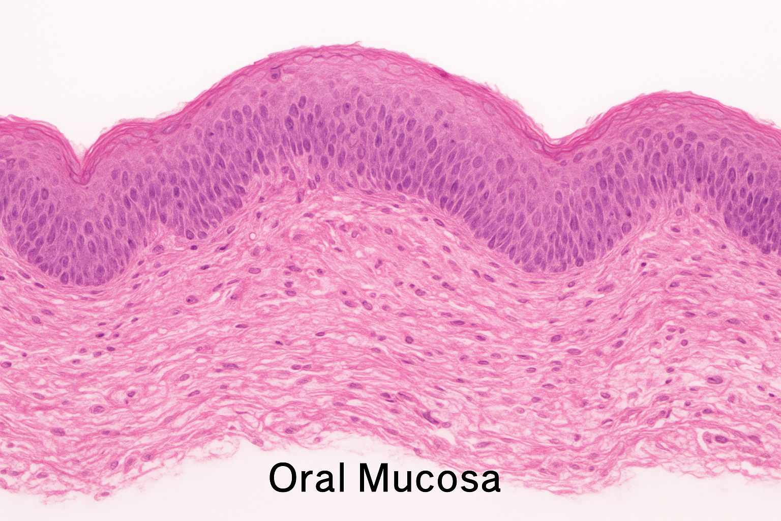
The oral mucosa consists of a stratified squamous epithelium, and a layer of connective tissue beneath the layer called lamina propria. In some areas, this mix of the tissue is anchored by submucosa and can include small glands of saliva, blood vessels and nerves. Oral mucosa has different histology throughout depending on the location, mechanical stress and need of the oral cavity leading to the formation of three different types:
- Lining mucosa
- Masticatory mucosa
- Specialized mucosa
The differences between these types include epithelial thickness, keratinization, the structure of connective tissue, and the overall resilience of these types which is correlated with adapting to certain functions of the mouth.
Lining Mucosa
Mucosa is lined on most of the oral cavity such as inside cheeks, lips, soft palate, floor of the mouth, and backside of the tongue. Its main functions include the ability to offer a flexible and protective cover that can withstand the slight mechanical forces yet remain mobile when speaking and chewing food.
Histological Features
- Epithelium Non-keratinized stratified squamous epithelium is the most common, although there are places where parakeratinization may occur. The epithelial layer is not so thick and it has less layers of cells than masticatory mucosa has.
- Lamina propria: Under the epithelium lies loose connective tissue with plenty of elastic fibres which give it mobility and cushions.
- Submucosa: It is found in most of the areas of lining mucosa and is composed of minor salivary glands, adipose tissue and blood vessels. This aspect makes lining mucosa less rigid.
Functions
- Gives it a loose lining to allow oral motions.
- Easy speech and swallowing.
- Provides defense against microbial invasion and other minor injuries.
Clinical Significance
- Lining mucosa is more vulnerable to traumatic damages like burns or lacerations because it is not keratinized.
- This tissue type is where common mucosal diseases such as aphthous ulcers, viral infections and oral candidiasis are located.
- Its histology knowledge is useful in the surgical planning, especially in the design of flaps and grafting, where flexibility and vascularity are important.
Masticatory Mucosa
The masticatory mucosa is adapted to resist the mechanical action of the chewing and mastication. It envelops the hard palate and the tooth gingiva, the areas that are under consistent pressure, friction and abrasion.
Histological Features
- Epithelial: Keratinized stratified squamous epithelium (which is either orthokeratinized (lacks nuclei in the top layer) or parakeratinized (preservation of nuclei). This keratinization gives me the added resistance to mechanical trauma.
- Lamina propria: This is dense connective tissue composed of collagen which is firmly attached to the periosteum of the underlying bone and provides structure and strength.
- Submucosa: It is frequently absent or of small amount, especially in places such as the attached gingiva thus making the tissue hard and immobile.
Functions
- Guards behind machinery when chewing.
- Supports the teeth and artificial appliances.
- Plays a role in sensory experience of pressure and pain in the process of mastication.
Clinical Significance
- The masticatory mucosa is rigged and makes it the most useful in dental practice like implant placement and periodontal surgery.
- Masticatory mucosa is prone to the manifestation of gingivitis and periodontal diseases, and therefore, accuracy in histological knowledge should be prioritized.
- The hyperkeratosis can develop in the response to chronic irritation of the keratinized epithelium which is less susceptible to ulceration.
Specialized Mucosa
The dorsal surface of the tongue has specialized mucosa which is adapted to gustation. This mucosa has taste buds and it is involved in sensory perception.
Histological Features
- Epithelial: The major part of the dorsal tongue is covered by epithelium which is a keratinized stratified squamous epithelium. The lateral and ventral surface is not keratinized.
- Papillae: There are several kinds of papillae (filiform, fungiform, circumvallate, and foliate), and each of them can have a different structure:
- Filiform papillae: Keratinized conical papillae, and the most abundant, basically mechanical in their action.
- Fungiform papillae: This is mushroom-shaped, and is lightly keratinized with taste buds.
- Circumvallate papillae: These are large, dome-shaped and located in the tongue at the back and hold a large number of taste buds.
- Foliate papillae: Taste buds are found on the lateral folds in the shape of a leaf.
- Lamina propria: Well-vascular connective tissue, which helps to provide the work of papillae and taste buds.
Functions
- Supporters taste sense with the help of specialized sensory structures.
- Offers a scuffed surface that helps with the handling of food.
- Contributes to the immune response of the mouth, as lymphoid tissue resides in some areas.
Clinical Significance
- Special mucosa can be lesioned or inflamed and impact on taste perception and oral sensation.
- Some general illnesses, including nutritional deficiencies or autoimmune diseases, are first identified in the mucosa of the tongue; as a result, histological knowledge is diagnostically essential.
- Surgery on the tongue must be done with knowledge of the layers of the mucosa to avoid the impairment of functions.
Comparison of Oral Mucosa Histology
| Feature | Lining Mucosa | Masticatory Mucosa | Specialized Mucosa |
| Epithelium | Non-keratinized | Keratinized | Keratinized or lightly keratinized |
| Lamina propria | Loose, elastic | Dense, collagen-rich | Rich vascularization, supports papillae |
| Submucosa | Present | Minimal or absent | Variable, under papillae |
| Location | Inner cheeks, lips, soft palate | Hard palate, gingiva | Dorsal tongue |
| Function | Flexibility, protection | Resistance to mechanical stress | Taste perception, food manipulation |
| Clinical importance | Ulceration, infection-prone | Periodontal disease, surgical base | Diagnostic indicator, taste disorders |
These differences are critical in understanding to initiate specific clinical interventions and disease control. As an example, oral surgeons should understand that grafts of lining mucosa are soft, and those of masticatory mucosa resist trauma.
Oral Mucosa Histology as a Clinical Application
Dentistry
- Proper understanding of the mucosa of the mouth can be used in the restorative dentistry, planning of the prosthetics, and the implantology.
- As an instance, keratinized masticatory mucosa offers a firm support to dental implants and bridges that may otherwise have a tendency of receding.
- It is advantageous to line mucosa with its elasticity when constructing flaps of periodontal surgery.
Oral Surgery
- The histological knowledge aids the surgeons in strategizing incision and grafts to reduce the postoperative complications.
- In case of reconstructive surgeries, tissue of the hard palate (masticatory mucosa) is usually used due to its durability.
- When conducting tongue surgeries, it is important to identify specialized mucosa that would retain the ability to taste.
Disease Diagnosis
- Numerous systemic and mouth diseases appear in certain types of mucosa.
- Aphthous ulcers, viral infections and candidiasis often impact on non-keratinized lining mucosa.
- The site of the leukoplakia and the keratotic lesions are found in the masticatory mucosa as a result of chronic mechanical irritation.
- Mucosa alterations can show some specialized alterations which can serve as evidence of nutritional deficiencies (e.g., atrophic glossitis) or autoimmune disease such as lichen planus.
Conclusion
The oral mucosa is a specialized tissue that is dynamic and exhibits three different types tailored towards certain functions in the mouth cavity. Mucosa lining gives it flexibility and protection, masticatory mucosa endures mechanical forces and specialized mucosa is the one that supports the experience of taste. This knowledge will be important in dentistry, oral surgery, and clinical diagnostics to understand their histological differences. Proper identification of these types of tissues aids clinicians to plan successful interventions, anticipate tissue reaction to trauma and detect pathological alterations in the initial stages.
Histology is not only a scholastic concept, therefore, it is an instrument that can be applied in practical patient care and clinical decision-making. Knowledge of the structure and functionality of the oral mucosa will improve the results of restorative, surgical, and diagnostic dentistry by making sure that the treatment is safe, effective, and meets the tissue-specific needs of the oral cavity.