The neck is a very complicated and crucial part of the human body. It helps to connect the head with the torso, giving it structural support, mobility and protection to important neurovascular structures. Knowledge about the anatomy of the cervical spine and support ligaments, muscles is important to health and fitness professionals as well as medical practitioners. This information not only points to the tight-rope between the flexibility and stability of the neck but also throws a new light on such widespread conditions as neck pain, whiplash injuries, and degenerative disorders.
Overview of the Cervical Spine
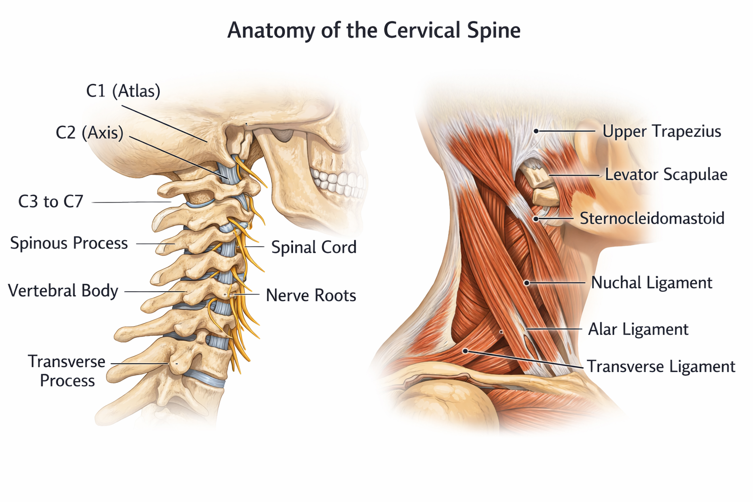
The vertebral column has the cervical as the topmost part, which comprises seven vertebrae that are identified as C1-C7. The vertebrae are specially shaped in such a manner that they can give both mobility and stability, which makes the head move in various directions, protecting the spinal cord and related nerve roots.
The atlas (C1) and axis (C2) are the first two cervical vertebrae which are unique to hold the skull and help the head turn about. The atlas holds the occipital condyles of the skull and thus allows nodding movements like yes, and the axis contains a projection of bone known as the odontoid process or dens that permits the head to swing side to side like the word no.
The other cervical vertebrae (C3-C7) are more orthodox, with vertebral body, pedicles, laminae, and spinous processes. The vertebrae create a column that is flexible and protective and that protects the spinal cord, vertebral arteries and the nerve roots and allows the movement of flexion, extension, bending laterally and turning.
Vertebral Anatomy of the Cervix
All the vertebrae of the cervix have distinct anatomical characteristics:
C1 – Atlas
- The vertical body is absent in the form of rings.
- Supports the skull.
- Gives the atlanto-occipital joint flexion and extension.
C2 – Axis
- Has the odontoid process (dens) which sticks out upwards.
- Bonds to the atlae.
- Allows rotation of the head.
C3-C6
- Normal cervical spinal vertebrae are small-bodied.
- Vertebral arteries have openings in transverse processes.
- Spinous processes tend to be bifid (divided) so that they can attach ligaments.
C7 – Vertebra Prominens
- Enlarged spinous process, which is palpated easily at the neck base.
- Elevates as an interspinal bone to the thorax spine.
The structure of the cervical vertebrae is focused on the lightness and flexibility of the vertebrae, as the bodies of the vertebrae are small, and the vertebral foramina are large, which ensures the motion of the neck and head without compromising safety of the spinal cord.
Cervical Spine Curvature
The cervical spine has a characteristic lordotic curve which is an inwards curve with the intention of spreading the mechanical stress during motion and carrying the weight of the head. This curve plays a very important role in ensuring balance, posture and effective operation of muscles and ligaments.
Poor posture, trauma, and degenerative changes may result in alterations of this curvature like a straightened or reversed cervical lordosis. These deviations can result in muscle tension, pain in the neck and compression of nerves.
Cervical Spinal ligaments
Ligaments are fibrous bands which are powerful and connected bones and hold the joints steady. The ligaments are important in the cervical spine because they regulate the movement and limit the excessive movement that may cause damage to the spinal cord or the vertebrae. The important ligaments of the cervix are:
Anterior Longitudinal Ligament (ALL)
- Traces on the anterior of the vertebral bodies.
- Avoids hyper extension of the neck.
- Maintains the anterior stability.
Posterior Longitudinal Ligament (PLL)
- This is situated in the vertebral canal at the posterior side of the vertebral bodies.
- Restricts hyperflexion.
- Helps to stabilize the spinal cord by preventing the intervertebral discs in the back.
Ligamentum Flavum
- Bonds laminae of the bones of the neighboring vertebrae.
- Elastic, as the spine bends and with the help of the spine straightens.
- Protects the spinal cord when there is a movement.
Interspinous and Supraspinous Ligaments
- Interspinous ligaments are inter-spinous process ligaments.
- Supraspinous ligament is positioned on the end of the spinous processes.
- Both limit too much flexion and give muscular attachment points.
Cruciate and Alar Ligaments
- Located at the sub-occipital area.
- The atlas and axis are held with the help of the cruciate ligament.
- The excessive rotation is prevented by the alar ligaments which safeguard the dens and the spinal cord.
These ligaments are important in terms of their integrity. Damage of cervical ligaments may cause instability, pain, and predisposition to spinal cord injury especially following trauma (whiplash).
The muscles that support the cervical spine are as follows. The cervical muscles are important as they help in the movement of head and neck, posture, and protection. These are grouped into anterior, posterior and lateral groups:
Anterior Neck Muscles
- Sternocleidomastoid (SCM): This is a run of the sterna and clavicle to the mastoid process. It allows the rotation and flexion of the head.
- Scalenes: The muscles are used lateral muscles that aid in flexing and lateral bending of the neck. They also raise the first two ribs during inhalation.
Posterior Neck Muscles
- Trapezius (fibers on the upper segment): Lifts up scapula and helps in the extension of the neck.
- Splenius Capitas and Splenius Cervicis: Strauss, turn head and neck and move it laterally.
- Semispinalis Capitas: Intense extensors and rotators of the cervix.
Deep Cervical Muscles
- Longus Colli and Longus Capitis: These are located in front of the vertebral bodies and they offer flexion, stabilization and maintenance of posture.
- Suboccipital Muscles: This is a small intricate muscle that attaches C1 and C2 to the skull, which is involved in fine motor control and head stability.
These are muscles, which interact with the ligaments to ensure stability of the neck as well as to enable fluid and controlled movement. Poor, tight, or damaged muscles are some of the causes of chronic neck pains, headaches as well as postural disorders.
Intervertebral Discs and Mobility of the Cervix
In between every cervical vertebra, there is an intervertebral disc, which consists of a soft, gelatinous nucleus pulposus and is surrounded by a tough fibrous annulus fibrosus. These discs are shock absorbers and help to move in between the vertebrae.
Intervertebral disc degenerative changes, including disc herniations and degeneration, may produce cervical nerve root or spinal cord compression resulting in such symptoms as neck pain, numbness, tingling, or arm weakness.
Blood Supply and Pathways of Nerves
The vital blood vessels and nerves pass through the cervical spine and serve the head, neck and the upper limbs:
- Vertebral Arteries: Proceed upwards through the transverse foramina of C1-C6 to create the basilar artery which feeds the brainstem and the posterior parts of the brain.
- Cervical Nerves (C1-C8): They exit through intervertebral foramen to serve muscles and skin of the neck, shoulders and upper limbs.
- Sympathetic Chain: This occurs laterally to the back of the vertebral bodies, and it affects the autonomic processes.
A blow or a squeeze of these structures can lead to the feeling of dizziness, headache, numbness or even neurological impairments.
Cervical Spine Disorders
The knowledge of the cervical spine anatomy can be used to identify the common conditions and their implications:
Cervical Spondylosis
- Vertebral degenerative alterations and disc degenerative alterations.
- Results in stiffness, pain and even compression of nerves.
- In most cases associated with wear and tear.
Whiplash Injuries
- Sudden trauma that causes hyperextension and hyperflexion like car accidents.
- Attacks the muscles and ligaments and at times the vertebrae too.
- The symptoms comprise headaches, stiffness and neck pain.
Herniated Discs
- The nucleus pulposus is displaced out of the annulus fibrosus.
- May cause compression of nerve roots and result in radiating pain, numbness, or weakness.
Postural Strain
- The sitting forward position of the head or bad posture.
- Pushes the cervical muscles and ligaments, leads to the development of chronic pains and tension headaches.
The timely diagnosis and adequate treatment with the help of physical therapy, posture training, and even surgical procedures can help people with cervical spine disorders to a considerable extent.
Caring for Healthy Cervical Spine
Good posture, strong muscles, and flexible ligaments are the attributes of a healthy cervical spine. Recommendations include:
- Frequent Strengthening of the neck and upper back muscles.
- Ergonomic Arrangement: Have the right desk and monitor height so that one is not leaning forward.
- Proper Sleep Position: Pillows to assist in the neutral position of the cervical position.
- Prevent repetitive Strain: Have a rest after spending time on the phone or computer.
- It should be evaluated early in the evolution of Medical Care: Deal with chronic pain, numbness, or weakness.
These measures prevent the risk of the injury and support the mobility and the functioning of the cervical spine during the aging process.
Conclusion
The cervical spine is a wonderful anatomical structure that maintains support and protection of the head and neck as well as its mobility. Its vertebrae, ligaments, muscles, intervertebral discs, and neurovascular structures all form a fine balance enabling the neck to move freely and prevent the spinal cord as well as protect the important functions.
Change of this balance, either due to trauma, degeneration or poor posture, may result in severe pain and disablement. Knowing the anatomy of the cervical spine, people will be able to appreciate the value of neck health, preventive measures and know when medical help is needed. A fit, supple body with good posture will be enough to have the cervical spine as stable, mobile, and protective well into the life span.