The human brain, despite making up a mere 2 percent out of the total body weight, absorbs almost 20 percent of all oxygen and glucose in the body. Such a high metabolic rate implies that the regular supply of blood is not only necessary to be normal but is also necessary and regulated so as to sustain the brain. Even the temporary failure of blood circulation in the brain can cause a loss of consciousness, irreversible brain damage, or death. The knowledge of the blood reaching the brain–of its protection–is, in consequence, an anatomical, physiological and clinical medical fundamental.
The key to this protection mechanism is the Circle of Willis which is a special ring of arteries at the bottom of the brain. This framework is extremely critical in ensuring cerebral perfusion, particularly in the instance where one of the major arteries is narrowed down or blocked. We are going to discuss the great arteries to the brain, study the anatomy and physiology of the Circle of Willis and relate the findings to all the key clinical conditions: stroke, aneurysms, and other cerebrovascular conditions.
Introduction to Cerebral Blood Supply
The brain has two main arterial systems, which pass in pairs ( one internal carotid artery and the other vertebral artery ) that supply it with blood. The existence of these vessels together makes sure that the blood is saturated with oxygen and is delivered to every part of the brain, including the cerebral cortex and the brainstem and cerebellum.
The Internal Carotid Arteries
The common carotid arteries give rise to internal carotid arteries on the neck. Having passed through the cervical area, each of the internal carotid arteries penetrates the skull via the canal of the temporal bone, carotid canal. When we get within the cranial cavity, these arteries give rise to a few important ones which nourish the anterior and middle parts of the brain.
- The internal carotid artery has major divisions namely:
- Anterior cerebral artery (ACA)- serves medial surfaces of frontal lobes and parietal lobes.
- Middle cerebral artery (MCA) – serves the lateral surfaces of the cerebral hemispheres such as parts that deal with motion, sensation and speech.
Posterior communicating artery- links the internal carotid system and the posterior circulation.
Carotid arteries are especially significant because they serve areas of the brain that are linked to higher order cognitive abilities, voluntary motor activities, and sensing ability.
The Vertebral Arteries
Vertebral arteries develop out of subclavian arteries and enter the cervical vertebrae through transverse foramina. They pass through the foramen magnum and converge at the bottom of the brain to make the basilar artery.
The vertebral and basilar arteries give branches to:
- The midbrain (brainstem, pons and medulla).
- The cerebellum
- The posterior sections of the cerebral hemispheres.
The posterior cerebral arteries (PCAs) are a major division of the posterior circulation and serve the occipital lobes and portions of the temporal lobes including portions of the temporal lobes involved in vision.
The Circle of Willis: Anatomical Structure
Circle of Willis: This arterial anastomosis is on the lower surface of the brain, and surrounds the optic chiasm and the pituitary stalk. It is a very important linkage between anterior (carotid) and posterior (vertebrobasilar) circulations.
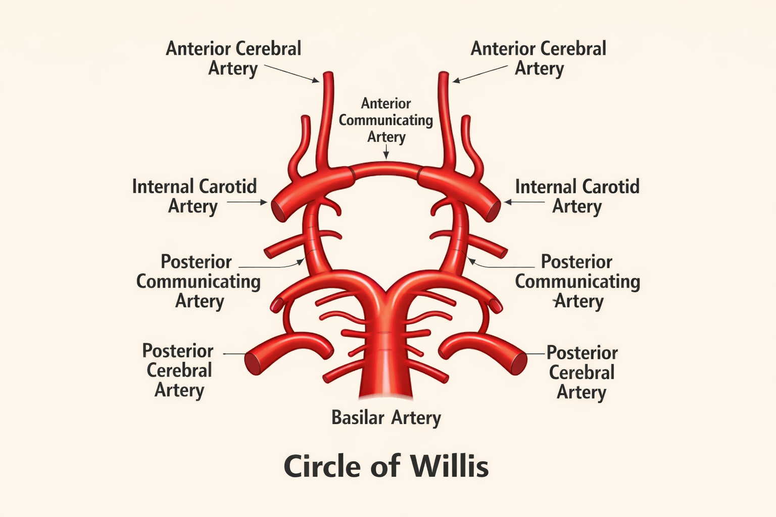
Elements of the Circle of Willis
The arteries that make the Circle of Willis are the following:
- The anterior cerebral arteries (left and right) are on the left and right sides respectively.
- Anterior communicating artery.
- internal carotid arteries (endings)
- On the right and left sides, there are posterior communicating arteries.
- Left and right posterior cerebral arteries.
These vessels make a fairly hexagonal ring together which enables blood to be reallocated among various sections of the brain in case of necessity.
Anatomical Variations
It should also be mentioned that a full Circle of Willis with all parts intact and well-formed can be seen only in approximately 20-25 percent of a population. Differences are usual and they might consist of:
- This is hypoplastic (underdeveloped) communicating arteries.
- Absence of certain segments
- Left and right vessel asymmetry.
Such differences may affect the regulating functioning of collateral circulation in the vascular compromise.
Functional Importance of the Circle of Willis
The main role of the Circle of Willis is to supply collateral circulation. This implies that it provides other avenues of blood circulation in the event that one of the great providing arteries is constricted or blocked.
Perfusion of the Cerebral Maintenance
In case a blood flow to one internal carotid artery is poor, there is the possibility to reroute blood through the anterior communicating artery of the other side. In the same way, the posterior communicating arteries enable blood in the vertebrobasilar system to feed anterior parts of the brain in case of need. It is a major protective aspect that contributes to the sustenance of sufficient oxygen supply to the neural tissue by this redundancy.
Pressure Equalization
The round structure of vessels also aids in equalizing the blood pressure in the anterior and posterior circulations. This minimizes the chances of unstable pressure that may cause rupture of frail cerebral vessels.
The Clinical Context of Circle of Willis
The Circle of Willis being a given understanding, interpretations of most neurological conditions and how to manage them are crucial. The structure and its role are directly connected to some of the largest cerebrovascular conditions.
Stroke and Ischemic Events
Stroke is a condition of the brain resulting in the cutting off of blood supply to a certain region of the brain due to blockage (ischemic stroke) or rupture of the vessels (hemorrhagic stroke). The Circle of Willis may occasionally compensate for the lower blood flow in ischemic strokes, which supply collateral circulation.
The well-developed Circle of Willis can lead to less acute symptoms or slower degeneration of neurological impairment in patients. On the other hand, the incomplete or poorly functioning circles are related to larger infarcts and a poor outcome.
Cerebral Aneurysms
Cerebral aneurysm is localized expansion of the blood vessel wall, which may turn up in the points of the arterial branches. The Circle of Willis often gets an aneurysm because of:
- High blood flow
- Vessel junctions hemodynamic stress.
- Weaknesses in the nature of arterial walls.
Ruptured aneurysm may result in a subarachnoid hematoma, a life-threatening event, which is marked by the onset of headache, neurological impairment and unconsciousness. The Circle of Willis anatomy anatomy is essential in the diagnosis and surgical treatment of such aneurysms.
Narrowing of the Vessels and Atherosclerosis
The atherosclerotic plaques may constrict the great arteries that serve the brain, especially the internal carotid arteries. Circle of Willis has the potential of sustaining blood flow in the brain at the onset of the illness. Nonetheless, in case of several vessels involved or when a circle is not complete, the risk of ischemia is very high.
Imaging and Diagnostic Relevance
Imaging and Diagnostic Relevance Imaging and diagnostic relevance tests are regularly conducted.
The cerebral circulation can now be visualized in detail using the modern methods of imaging. Common modalities include:
- CT angiography (CTA)
- Magnetic resonance angiography (MRA)
- Digital subtraction angiography (DSA).
These instruments can be used to determine the anatomy of the Circular of Willis, and any variation, aneurysms and the status of collateral blood flow. This kind of information is crucial in the planning of neurosurgical or endovascular.
Developmental and Evolutionary Perspectives
In the developmental perspective, the Circle of Willis is created in the early embryonic life when cerebral vessels differentiate and join. The anatomic variations of the adult body tend to show the embryological variation. Evolutionarily, having collateral pathways points to the role of safeguarding the brain against ischemic damage because it is the core of survival and behavior.
Conclusion
Blood flow to the brain is a highly sensitive and well-guarded mechanism that is aimed at coping with the tremendous metabolic requirements of the organ. The internal carotid and the vertebral arteries collaborate to ensure that blood flow in all parts of the brain is maintained with a supply of oxygenated blood. The core of this system is the Circle of Willis that is an arterial ring that is redundant, balances pressure and collateral circulation.
The anatomy and physiology of the Circle of Willis is not only critical to learners in the field of anatomy and physiology, but also to clinicians who handle strokes, aneurysms, and other cerebrovascular disorders. Its location could be a minor impairment to the neurons, or it could be a brain killer. The relationship between the anatomy and use of this knowledge in clinical practice is a potent illustration of how structure and function are intertwined in the human body by the example of the Circle of Willis.