The salivary glands are very important parts of the oral cavity, they help in the production of saliva (a fluid that facilitates digestion, lubrication, antimicrobial protection and all the overall homeostasis of the mouth). Knowledge of the histology of salivary glands is essential in understanding how saliva is generated, the functioning of the glands and how structural or cellular alterations can be the cause of oral diseases like xerostomia (dry mouth), bacterial or viral infections and autoimmune diseases like Sjogren syndrome.
The article will discuss the microscopic structure of the major and minor salivary glands, explain how saliva is produced physiologically and how histological issues can impact oral health.
Overview of Salivary Glands
There are two types of salivary glands in human beings:
Major Salivary Glands
They are paired and large (external to the oral mucosa) glands that lead to ducts that drain into the mouth. They are the ones that secrete most of the saliva.
- Parotid glands
- Submandibular glands
- Sublingual glands
Minor Salivary Glands
These are many small glands distributed all over the mouth mucosa, such as lips, cheeks, palate, tongue, pharynx etc. Their salivation is permanent to ensure the mucosal humidity.
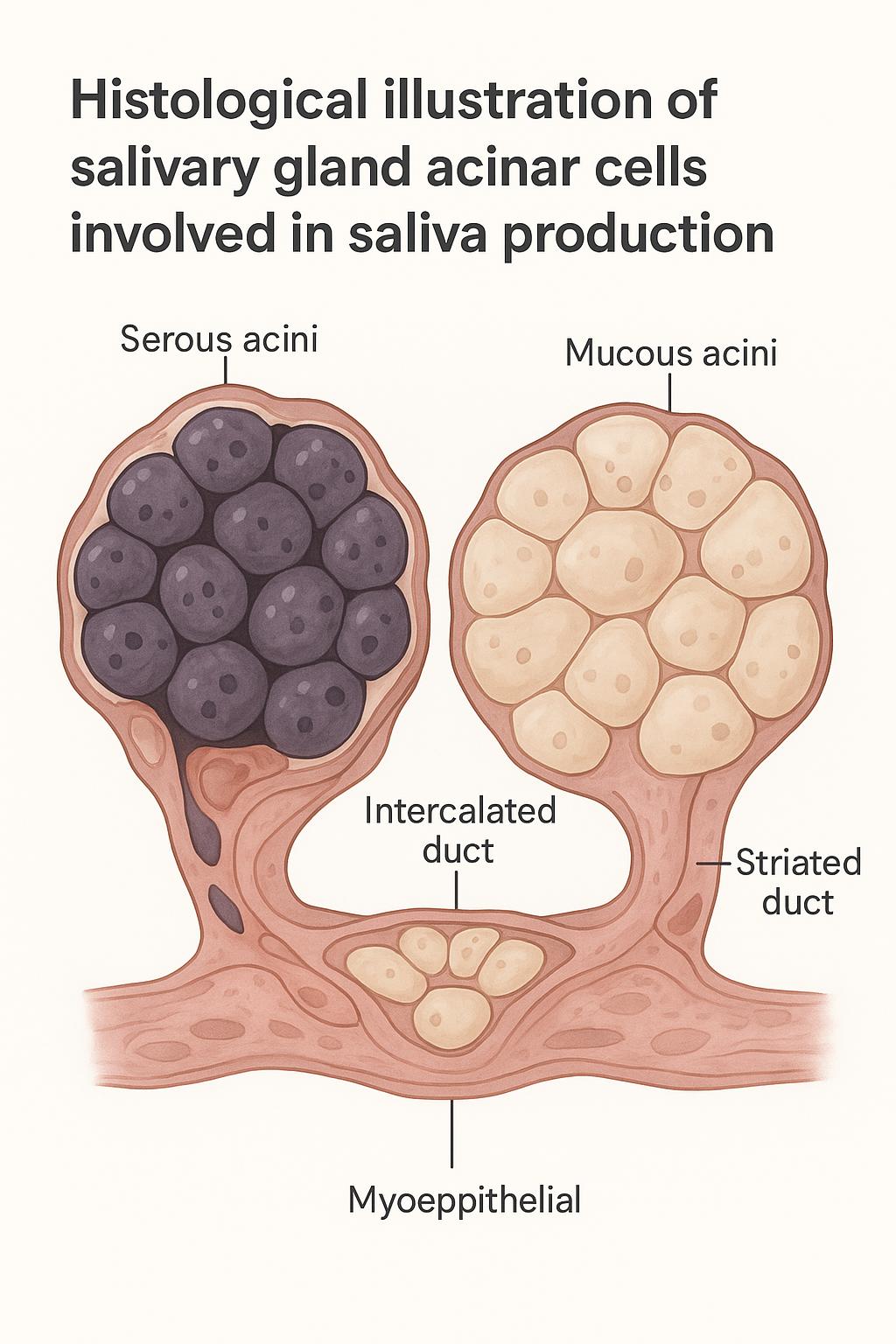
Salivary glands are histologically made up of secretory units, the acini, secretion conveying ducts, and supporting connective tissues. The organization and type of cells in each gland determines the make up of the saliva produced by it and its functions.
Microscopy of Salivary Glands of Major Size
Parotid Gland Histology
The largest of the salivary glands is the parotid gland which is solely serous.
Key histological features:
- Serous acini: round-naked pyramidal cells with basophilic cytoplasm as a result of high levels of rough endoplasmic reticulum.
- Zymogen granules: appear at the apical level, including amylase – an enzyme necessary in the initial step of the digestion of carbohydrates.
- Large ductal system: There are intercalated ducts and striated ducts of the parotid.
Functionally:
- Sews watery saliva that is enzyme rich.
- This plays a crucial role in digestion, especially breaking down of starch.
The Histology of the Submandibular Gland
Submandibular gland is a mixed gland composed mostly of serous acini.
Histological characteristics:
- Mixed acini: mucous acini are topped by serous cells in the shape of crescent (serous demilunes).
- Striated ducts: these are well developed and necessary to adjust the electrolyte composition.
- Secrets approximately 60-70 percent of the rest saliva.
Functional outcomes:
- secretes slightly thick saliva.
- Significant source of resting saliva, which keeps oral mouth moist.
2.3 Sublingual Gland Histology
The sublingual gland is mostly mucous.
Key structural attributes:
- Most acini are mucous acini, which adds to the thick gel-like secretions.
- There are serous demilunes although not overpowering.
- The ducts are underdeveloped and unlike parotid and submandibular glands.
Functional implications:
- Secrets mucus laden saliva, which is essential in lubrication.
- Aids in speech, swallowing and protection of the mouth.
Minor Salivary Glands Histology
Most of the salivary glands found in the mouth are mucous, with exceptions of serous glands found in the von Ebner’s glands close to the circumvallate papillae.
Microscopic features:
- Clusters of mucous acini.
- Short, poorly defined ducts.
- Imbibed in the connective tissue in the oral mucosa.
Importance:
- Have constant lubrication of the base.
- S secrete saliva that is high in IgA, mucins and antimicrobial proteins.
These glands are very important in ensuring that the body does not dry up particularly when sleeping, when the major salivary gland activity is at a low level.
Process of Saliva Production
The production of saliva starts in the acinar cells and the composition of the saliva is altered as it passes through the ductal system.
Acinar Cell Secretion
- Serous cells produce a liquid fluid, which is filled with enzymes (e.g., amylase, lipase).
- On the mucous cells, mucins are secreted and hydrated to be mucus.
Ductal Modification
Modification of the original fluid is done by the duct system:
- Added bicarbonate is by intercalated ducts.
- Striated ducts modify ions:
- Absorb sodium and chloride
- Secrete potassium
This process would make sure that saliva contains a proper pH, electrolyte makeup and viscosity to sustain the health of the mouth.
Final Saliva Composition
Final saliva contains:
- Digestive enzymes
- Mucins
- Electrolytes
- Antimicrobial peptides
- Immunoglobulins (particularly, IgA)
This mixture promotes digestion, lubrication, buffering, as well as immune defense.
Functional Roles of Saliva
Lubrication and Moisture
Mucins attract water and maintain the moisture of the tissues of the mouth and avoid tissue damage.
Digestion
Amylase among other enzymes start the digestion of carbohydrates in the mouth.
Immune Response towards Pathogens
Saliva contains:
- Lysozyme
- Lactoferrin
- Defensins
- IgA
These elements prevent the growth of micro-organisms.
pH Buffering
Bicarbonate ions counteract acid and save the enamel of teeth.
Tissue Cleansing
Saliva keeps depositing garbage and bacteria.
Histology and Its Effects on Oral Health
Any harm or change in the histology of the salivary glands causes a deficiency in the production of saliva, which causes severe oral health issues.
Xerostomia (Dry Mouth)
Causes:
- Atrophy of acinar cells
- Radiation therapy damage
- Drugs (antihistamines, antidepressants)
- Blocking of ducts (e.g., of stones)
Histological Findings
- Loss of acinar cells
- Fibrosis (substitution with connective tissue)
- Chronic infiltrates inflammation.
Consequences:
- Difficulty swallowing
- The risk of dental caries increased.
- Oral (candida overgrowth) infections.
- Cracked mucosa
Salivary Gland Infections
Bacterial Infection (e.g., sialadenitis)
Often due to salivary stasis.
Histology:
- Neutrophilic infiltration
- Ductal dilation
- Suppuration
Symptoms:
- Pain
- Swelling
- Fever
Viral Infections (e.g., mumps)
Affect mostly the parotid glands.
Histology:
- Acinar cells degeneration.
- Edema
- Lymphocytic infiltration
Outcome:
- Decreased salivation in case of infection.
- Possible atrophy of the glands in the long-term.
Autoimmune Disorders Sjogren Syndrome.
Sjogren Syndrome is an autoimmune disorder of the body, affecting the body’s replacement cells and glands (Gottlieb 2019).
Sjogren syndrome is considered to be one of the most important diseases of salivary gland histology.
Mechanism:
The immune system attacks exocrine glands, particularly salivary and lacrimal glands.
Histological hallmarks:
- Foci of lymphocytic infiltrations.
- Destruction of acinar cells
- Ductal hyperplasia of epithelia.
- Substitution of either gland or fat tissue with fibrous tissue.
Clinical manifestations:
- Severe dry mouth and eyes
- Disability in speaking and swallowing.
- Increased dental decay
- Oral fungal infections
This knowledge of these histological patterns is critical in the diagnosis of Sjogren syndrome as well as the progression of the disease.
Salivary Glands- Histology Clinical Significance
History Salivary glands, when examined histologically, give important diagnostic data.
Key diagnostic uses:
- Determination of the causes of persistent dry mouth.
- Hypersigns of salivary gland tumors.
- Diagnosing autoimmune diseases.
- Evaluating inflammatory diseases.
- Assessment of radiation tissue damage.
The results of the biopsy inform treatment options including drug therapy to stimulate saliva secretions and immunosuppression.
Cleaning Healthy Salivary Glands
Even though certain factors that lead to dysfunction of the salivary glands cannot be avoided, there is a range of preventive measures, which could help to preserve the well-being of the glands:
- Stay hydrated
- Have a high level of oral health.
- Restrict alcohol and caffeine (which decrease saliva)
- Manage systemic diseases
- Take lozenges or sugar-free gum to help in stimulating saliva.
- Avoid smoking
- Request immediate attention to infections.
Such habits ensure the maintenance of the structure and functioning of glands.
Conclusion
Histological study of salivary glands gives an in-depth explanation of how the organs generate saliva and keep the mouth healthy. The serous-dominant parotid gland has its way through to the mucous-filled minor glands which have their own distinct functions in regard to digestion, lubrication, immune defense, and tissue maintenance.
The reduction of salivation production results in severe oral and systemic complications when histological alterations take place, such as infection, autoimmune disease, radiation, aging, etc. The identification of these microscopic patterns assists clinicians to diagnose and treat the following conditions such as xerostomia, sialadenitis and Sjogren syndrome.
Knowing the close relationship between glandular histology and oral health will enable clinicians and patients to take the initiative to ensure that they maintain a functioning salivary system.