The process of tooth development, also referred to as odontogenesis, is a complicated and tightly regulated process, which starts at a very early stage during embryonic life and goes on until the teeth emerge into the oral cavity. It is a complex chain of interactions among genetic, molecular and environmental factors which coordinate the growth shape and differentiation of dental tissues. The study of tooth formation stages offers a good perspective on the formation of teeth, why some people have dental abnormalities and why some teeth erupt sooner than others.
Introduction: Anatomy of Tooth Formation
Development of teeth is not a biological accident, but a specific physiological process that starts approximately in the course of the sixth week of embryonic development. Cells in the oral epithelium and underlying ectomesenchyme at this point react to start the tooth germ. The special types of cellular activities of each stage of development, including initiation, morphogenesis, histodifferentiation, and eruption, determine the final structure and activity of the tooth.
Mechanical digestion, speech, and aesthetics of the face all depend on teeth. Their growth is a function of an interactive relationship between genetic directions and environmental factors. Violations of this process can result in the absence of teeth, malformation or slow eruption.
The Early Phase: Tooth Development
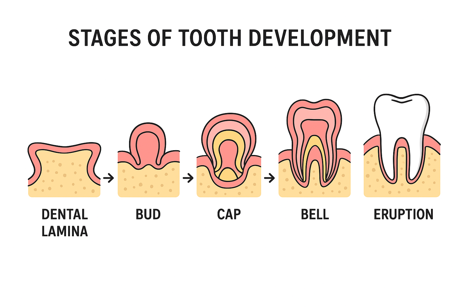
The tooth formation starts in the initiation stage. It is during the sixth week of intra-uterine existence that the oral epithelium becomes thickened along the eventual dental arches to create the primary epithelial band which subsequently separates into two structures- dental lamina and the vestibular lamina.
- Dental Lamina: This is the structure that becomes the base of the tooth bud of the primary and later, of the permanent dentitions.
- Vestibular Lamina: This is the space that occurs between the teeth and the inside of the cheeks or lips.
This phase depends on the epithelial-mesenchymal cross-interactions greatly. Where and when teeth buds are to form, is dictated by molecular signals, in particular those that deal with growth factors like FGFs (Fibroblast Growth Factors) and BMPs (Bone Morphogenetic Proteins).
There is a huge role of influences of genetics. Gene mutations that cause hypodontia (absence of teeth), supernumerary teeth (extra teeth), can be caused by hyperactivation of particular signaling pathways and PAX9 and MSX1 are the genes involved.
The Bud Phase: The Immortality of the Tooth Germ
The bud stage starts when the dental lamina turns inward into the mesenchymal region to produce small outgrowths of the epithelial cells known as tooth buds. To every bud is a tooth to come.
At this level, the tooth germ is predominantly made up of the proliferation of epithelial cells and little morphologic differentiation. The mesenchymal cells surrounding the epithelial bud condense to form the basis of the dental papilla and dental follicle precursors to dentin, pulp, cementum and periodontal ligament.
Cell proliferation during this stage can be influenced by environmental influences (maternal nutrition, infections, teratogens (exposure to some drugs or radiation) and this may cause an abnormal size or number of teeth to be formed.
The Cap Stage: Cap first begins Tooth Shape Development
The cap stage is the beginning of morphogenesis, or the stage at which the shape of the tooth begins to develop. The epithelial bud folds further and enlarges to form a cap-like structure that covers a section of the dental papilla, the enamel organ.
The tooth germ comprises the enamel organ, dental papilla and dental follicle. The role of each of the components is significant:
- Enamel Organ: Produces ameloblasts which are the cells involved in the formation of enamels.
- Dental Papilla: This forms into odontoblasts (dentin forming cells) and the dental pulp.
- Dental Follicle: It forms the supporting tissues (cementum, periodontal ligament and alveolar bone).
The development of the shape of the crown including whether it will be a molar, premolar or incisor is determined by the signaling between these three layers. It is guided by the presence of the particular gene expressions such as the BMP4 and FGF8 gradients along the dental epithelium.
Interruption at this stage may result in malformation of tooth shapes like incisors in the form of a peg or taurodontism.
The Bell Stage: Long division and Tissue differentiation
The bell stage is a very important stage of histodifferentiation and morphodifferentiation. On differentiation of the epithelial cells, the enamel organ develops the bell shape with formation of specific layers:
- Inner Enamel Epithelium (IEE): The cells develop further to be ameloblasts which compose enamel.
- Outer Enamel Epithelium (OEE): Coats the forming enamel organ.
- Stellate Reticulum: This is a type of support that gives ameloblasts nourishment and support.
- Stratum Intermedium: The works that have ameloblasts during enamel mineralization.
In the meantime, the cells of the dental papilla undergo differentiation into odontoblasts and begin to secrete predentin which is subsequently mineralized to form dentin. When the dentin formation commences, the ameloblasts start to produce enamel.
Such a synchronized secretion also guarantees that the junction between dentino-enamel (DEJ) is fully aligned – a crucial structural joint of tooth strength.
Genetic mutations in enamel or dentin-related enamel and dentin-related genes like AMELX, DSPP, or ENAM could result in amelogenesis imperfecta or dentinogenesis imperfecta which involve defects in mineralization.
CrownFormation and Root Development
Following the bell stage, the body of the tooth is formed as enamel and dentin are secreted in layers and at rates that are genetically determined. When crown formation is finished, root formation is initiated by Hertwig’s epithelial root sheath (HERS).
HERS determines the root shape, number and length via the differentiation of root odontoblasts by stimulation of these cells. The sheath disintegrates later, which permits cementoblasts to produce cementum and bind the tooth inside the alveolar bone.
Abnormalities in HERS and associated signaling pathways may result in root dilaceration, fusion anomalies, or concrescence (cemental fusion of adjacent teeth).
Tooth Eruption: Hidden Germ to Functional Tooth
The eruption stage is the last and most apparent stage of tooth formation. Its definition is the movement of the developing tooth from within the alveolar bone into the oral cavity, where it starts functioning.
Tooth eruption occurs in three stages:
- Pre-eruptive phase: During development and growth within the jawbone, the tooth germ migrates.
- The Eruptive Teeth penetrate the gingiva and erupt into the oral cavity.
- After The Post-Eruptive Phase The tooth adapts its position even after eruption, during jaw growth and wear.
The process of eruption is influenced by multiple factors:
- Signaling from the dental follicle, in particular involving colony-stimulating factors to upregulate bone resorption where the tooth is emerging.
- Root elongation, periodontal ligament remodeling, and vascular pressure also contribute.
Genetic and Environmental Effects on Tooth Development
The rate and success of tooth development is influenced by genetic and environmental factors.
- Genetics:
MSX1 and PAX9 gene variants can influence tooth number as well as shape. For instance, agenesis of premolars and/or third molars is frequently due to MSX1 mutations.
- Environmental Factors:
Prenatal contact with toxins (such as alcohol, lead, or certain medications), inadequate maternal nutrition (particularly calcium and vitamin D deficiency), and young childhood diseases. can disrupt normal odontogenesis.
- Hormonal Influence:
An association of EPE with endocrine diseases (hypothyroidism or hypopituitarism) has been known.
Etiology and Clinical Consequences of Delayed Tooth Eruption
Late tooth eruption is a frequent developmental abnormality that may have genetic, systemic, or local causes. Common causes are:
- Inherited diseases: cleidocranial dysplasia and Down syndrome.
- Genetic diseases: Rickets, malabsorption syndromes and endocrinopathies etc.
- Local factors: Crowding in mouth or early loss of baby teeth or cystic lesions blocking the path of eruption.
Having a perspective on the physiological timeline enables the clinicians to determine whether a delay represents a normal variation or the presence of a pathological condition.
Developmental Origins of Dental Anomalies
As each phase of tooth development is tightly regulated by signaling between and within cells, perturbations can result in anomalies of the tooth. Examples include:
- Anodontia or hypodontia: Arrest of the developmental process in the stage of initiation with resulting absence of teeth.
- Supernumerary Teeth: Overactive initiation of tooth development generating more than the usual number of tooth buds.
- Microdontia or Macrodontia: Defective morphogenesis producing sized anomalies.
- Deficient activity of ameloblasts during histodifferentiation or calcification.
- Fusion or Gemination: Disturbances in morphogenesis which make two tooth germs join or one tooth germ partially divide.
Not only do these irregularities present challenges to dental aesthetics, but they also pose risk to occlusion and oral function, thereby necessitating orthodontic and/or restorative care.
In summarizing…the miracle of odontogenesis
The development of the tooth from the bud stage to eruption seems to be one of the most synchronized events in human nature. This is particularly true for the phases of initiation, morphogenesis, histodifferentiation, and eruption, each of which depends on a precisely balanced interaction of genes, cellular signalling and environmental factors.
An advanced understanding of the stages of tooth development enables not only the diagnosis and management of developmental dental anomalies but also provides the basis for advancements in regenerative dentistry, stem cell therapy, and biomimetic enamel engineering.
In the end, the growth from a microscopic cluster of cells to a whole functional tooth is an extraordinary testament of biological precision and resilience — one that will surely continue to fuel research in regeneration and repair of dental tissues.