Oral cancer is one of the major health challenges in the world, and thousands of fresh cases are made annually. The early detection is essential to enhance the patient outcomes and survival rates, but the disease is usually not noticed until it is in the advanced stage. The microscopic study of a tissue, which is known as histopathology, has been central in the detection of precancerous lesions and early oral cancer. The knowledge of the processes, steps and advantages of histopathological analysis will enable clinicians to act promptly and provide relevant treatment.
Understanding Oral Cancer
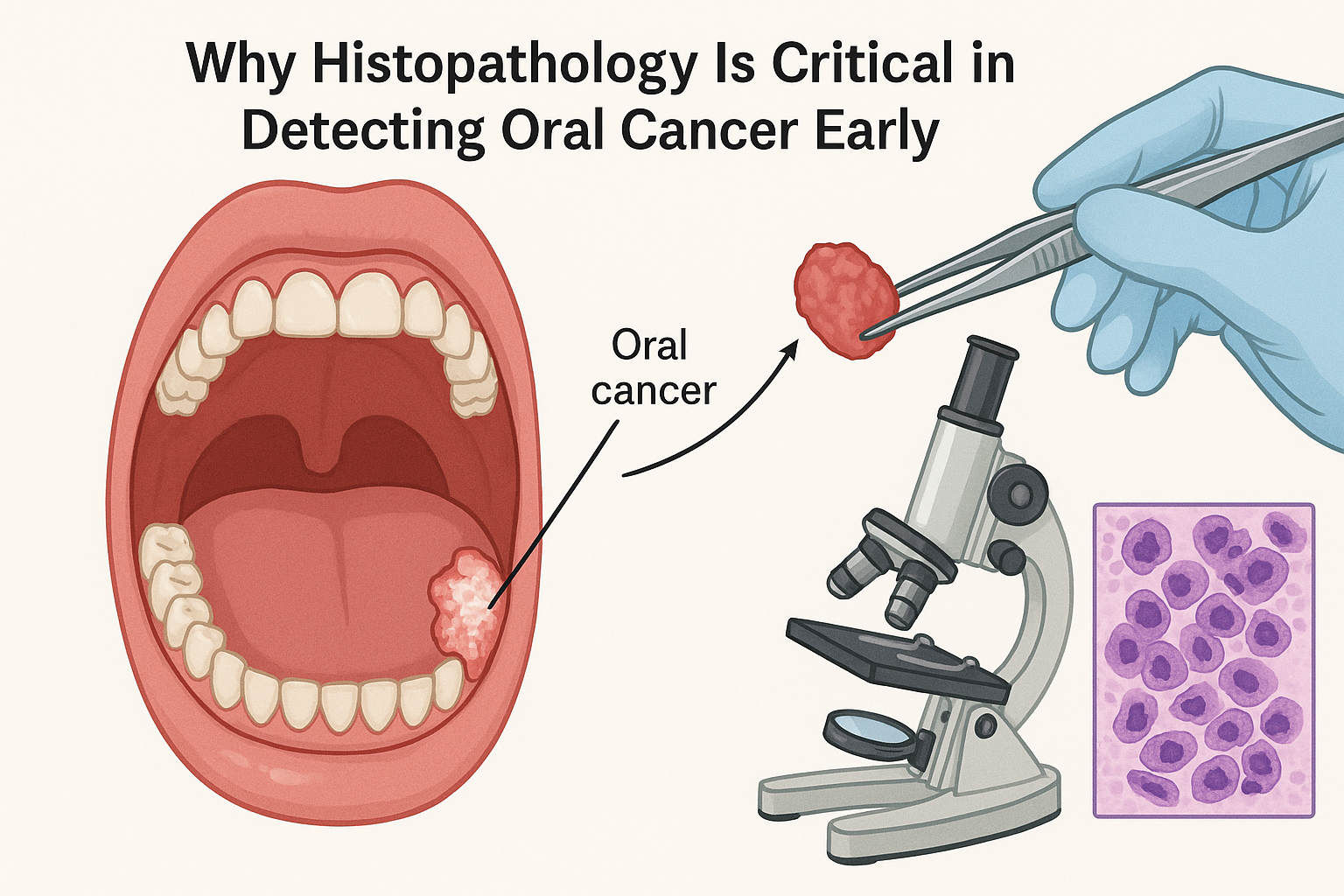
Oral cancer is malignant growths which develop in the tissues of the mouth such as the lips, tongue, floor of the mouth, buccal mucosa and palate. The most prevalent one is squamous cell carcinoma that starts with the epithelial lining of the mouth cavity. Tobacco use, alcohol abuse, human papillomavirus (HPV) infection, and chronic irritation of ill-fitting dentures are examples of risk factors.
The early-stage oral cancer disease has subtle symptoms that include small white/red spots, non-healing ulcers or slight edema. The symptoms can be easily confused with other benign conditions, thus, diagnosis can be delayed due to the use of clinical examination. The invaluable thing in this is histopathology.
The Purpose of Histopathology in Early Detection
Histopathology is the study of the architecture of tissues and cellular alterations using microscopy. With the help of a microscope, pathologists have the chance to observe abnormalities in biopsied tissue, which points to malignancy even before any clinical signs can be seen. This accuracy enables detection of precancerous lesions which are disorders like leukoplakia, erythroplakia and oral submucous fibrosis that could become cancerous in case they are not allowed to heal.
Key Histological Markers
There are some cellular and structural alterations as red flags of early malignant transformation:
- Dysplasia- abnormal growth and differentiation of the epithelial cells. Dysplasia is graded as mild, moderate or severe and usually may lead to invasive carcinoma.
- High cytoplasmic ratio – The irregular enlarged nuclei are evidence of cell atypia.
- Destruction of normal stratification – The disturbance of the stratification of the epithelial cells may indicate the emergence of neoplastic changes.
- Hyperchromatism – Stains dark, as a result of heightened DNA content, which is indicative of cell proliferation.
- Mitotic figures – When abnormal mitosis is found, it is an indicator of a high cell turnover, which may be malignant.
The markers are seen under high magnification, and they are objective to differentiate the benign, premalignant, and malignant conditions, which are used in clinical decision-making.
Biopsy Procedures: Portal to Truly Diagnosing
The basis of histopathological diagnosis is a biopsy. It entails taking away of a small piece of tissue material to a suspicious site, then processed and examined by means of a microscope. It has a variety of biopsy procedures in the field of oral pathology:
- Incisional Biopsy
An incisional biopsy is used when the lesions are big or diffuse and a representative part of the lesion is excised. This can be used to provide successful diagnosis without affecting the integrity of the surrounding tissue.
- Excisional Biopsy
In case of small lesions, the whole lesion may be excised and characterized. Excisional biopsies are mainly diagnostic as well as therapeutic particularly in early-stage oral cancer.
- Punch Biopsy
A circular blade removes a core of tissue, which is usually used in mucosal lesions. Punch biopsies are less invasive and have the potential of giving sufficient tissue to undergo histopathological analysis.
Anchor link to more information: To find specific information on how to diagnose and treat oral cancer, go to Cleveland Clinic Oral Cancer Resource.
Improving Patient Outcomes through Microscopic Analysis
Histopathology is not just a confirmation of finding, but it has a direct impact on patient care. The early identification through tissue analysis has a number of vital benefits:
- Individualized Treatment Planning
The level of dysplasia/tumor differentiation identified according to histopathology assists clinicians to choose the best form of treatment. Oral cancer at an early stage can be treated using local excision, laser therapy, or limited radiation and the morbidity is minimized.
- Prognostic Information
Histological grading gives an idea of the probable behavior of the lesion. Poorly differentiated tumors may only be treated with aggression, whereas well-differentiated tumors are slower growing and have better prognoses.
- Monitoring Recurrence
Histopathology after the treatment enables the pathologist to evaluate the surgical margins, which is important to make sure that all malignant tissue has been removed. This decreases the chance of relapse and educates follow up.
- Recognizing Risky Patients
Monitoring programs can be enrolled with patients with precancerous lesions identified on a histological level. Repeat biopsies and frequent examinations can help identify the manifestation of malignant transformation at the very first stage possible.
State of Art Methods that improve Histopathology
There is modern development of pathology that has moved past the old methods of staining and has enhanced diagnostic accuracy of oral cancer:
Immunohistochemistry (IHC)
The IHC involves the detection of malignant transformation-related proteins through antibodies. Examples include overexpression of p53 or Ki-67, which is a good prognostic data of cellular proliferation.
Molecular Diagnostics
The genetic mutations and chromosomal abnormalities are identified using the techniques of polymerase chain reaction (PCR) and fluorescence in situ hybridization (FISH). These procedures supplement histopathology especially in indistinct ones.
Digital Pathology
With the help of high-resolution digital scanning and artificial intelligence (AI) algorithms, the pathologists are able to measure tissue patterns with higher precision and speed. This is able to minimize human error and harmonize diagnosis in institutions.
Benign Proliferation of Oral Mucosa
Clinical knowledge of the histological characteristics of precancerous lesions can be used to target early intervention:
- Leukoplakia – White spots, hyperkeratotic with epithelial dysplasia. Histology defines the threat of malignant development.
- Erythroplakia – Red velvety lesions that have a tendency of being linked to severe dysplasia or carcinoma in situ. Requires immediate biopsy.
- Oral Lichen Planus – This is a chronic inflammatory disease which is subject to malignant change, especially the erosive type.
- Submucous Fibrosis – The fibrotic changes of the mucous of the oral cavity associated with the chewing of the areca nut; histologically epithelial atrophy and dysplasia are seen in severe cases.
Early biopsy and histopathological analysis of these lesions can help greatly to cut down invasive oral cancer.
Problems of Histopathological Diagnosis.
Though histopathology cannot be overestimated, it has its limitations:
- Sampling Error – A small biopsy could not reveal regions of high-grade dysplasia.
- Subjective Interpretation – Dysplasia grading needs skills and inter-observer errors may arise.
- Resource Limitations – Some areas may have a lack of access to skilled pathologists and other high-end diagnostic equipment.
Although this is the case, the limitations of histopathology have many limitations as compared to its advantages of early detection.
The use of Histopathology in Routine Oral Screening
Frequent oral check-ups, together with low biopsy threshold, are essential in cancer diagnosis at an early stage. Dentists, oral surgeons and primary care providers ought to be educated to identify suspicious lesions and refer patients as soon as possible to histopathological examination. There can also be the thought of a public awareness campaign encouraging signs of oral cancer in early stages thus increasing the rate of early detection.
Conclusion
Early diagnosis and treatment of oral cancer cannot be done without histopathology. It enables clinicians to treat precancerous lesions and small cellular changes prior to development of invasive disease because it is capable of detecting them at an early stage. The most important histological markers, accurate biopsy methods, and novel methods of diagnosis all assist in enhancing patient results.
Tissue analysis creates an opportunity to identify early and not only increase the survival rates but also provide less aggressive treatment, saving the functionality and quality of life. Embarkation of histopathology into mainstream clinical practice is crucial in fighting the worldwide morphine of oral cancer and facilitate the provision of effective therapy to the patients in time.