Healthcare nowadays depends so much on imaging technology to diagnose, prepare and follow up on various medical conditions. In everyday check-ups, as well as in complicated operations, medical imaging is critical in helping to make proper diagnosis, treatment, and encouraging better patient outcome.
Offering a closer look at the inner organs, these technologies enable medical workers to identify anomalies, monitor the disease course, and assess the efficacy of treatment with the most accurate precision in the history of medicine.
Knowledge in Medical Imaging Technology
Medical imaging is a collection of processes used to produce visual images of the inside of a body to be analyzed in a clinical setting and medically operated. Comparing to the traditional physical examinations, imaging allows physicians to view tissues, organs, and skeletal structures without the need to be invasive. Imaging technologies have also made diagnostic medicine to become faster, safer and more accurate due to the development of imaging technologies in the last decades.
The commonest imaging modalities are X-ray, ultrasound, computed tomography (CT) and magnetic resonance imaging (MRI). All the modalities have their own physical principles and offer a variety of information concerning the body. Such methods are important to understand in order to value their clinical value.
X-ray Imaging
Principles of X-ray Imaging
One of the oldest and most typical types of medical imaging is the x-ray imaging. It operates by x-rays of the body. Since dense structures like bones absorb more X-rays they appear white on the resultant images and soft tissues are permeable to the X-rays hence appearing dark.
Clinical Applications
Bone fractures, infection, tumor, and dental issues are readily identified by use of X-rays. Moreover, there are special X-ray methods, including mammography that enable early diagnosis of breast cancer and hence, survival rates are high.
Benefits and Limitations
The X-ray imaging has the following great benefits: speed, accessibility and cost-effectiveness. Nonetheless, there are possible risks associated with the use of ionizing radiation particularly when used repeatedly. Thus, X-ray imaging is mainly used in the instances when the advantages exceed the risks.
Ultrasound Imaging
Principles of Ultrasound

Ultrasound imaging is the technique that uses sound waves of high frequency in order to create real-time images of soft tissues. Sound waves are emitted into the body by a transducer and it is reflected back on the tissues and sent to the device, forming a visual image of the internal structures.
Clinical Applications
The application of ultrasound in obstetrics includes fetal monitoring, cardiology, in blood flow and heart functions. One can also use it in imaging of the abdomen to analyze the abdominal organs such as the liver, kidney, gallbladder, and so on.
Advantages of Ultrasound
Ultrasound is non-invasive, ionizing radiation-free and provides real-time imaging, therefore, it can be used safely on a regular basis and to check on the current conditions. It is also portable and can also be used to examine the bedside in emergency care.
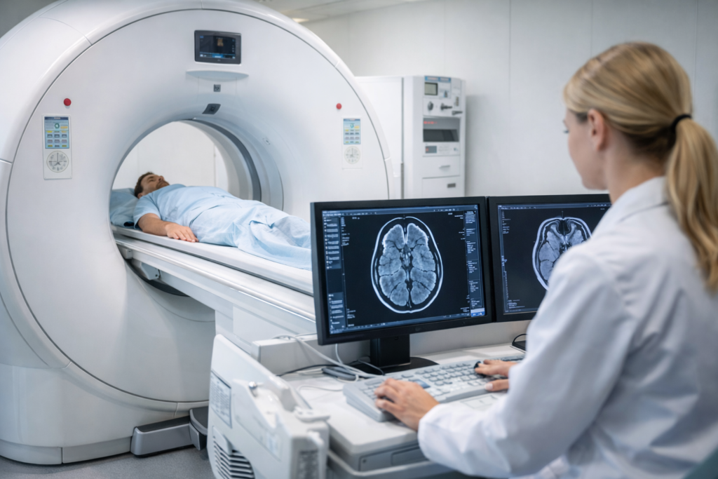
Computed Tomography (CT) Scan
Principles of CT Imaging
CT scanning is a process that involves the X-ray provided at different angles and then cross-sectional images of the body are obtained. Such measurements are now reconstructed into three-dimensional images and details by advanced computer algorithms enabling the visualization of the bones, organs and blood vessels with great precision.
Clinical Applications
The CT scans play a vital role in identifying vascular diseases, internal injuries, cancer, and neurological disorders. They can be used in emergency medicine particularly to assess cases of trauma at a glance.
Advantages and Reflections
Detailed anatomical data that is better than the conventional X-rays is offered by CT imaging. But similar to X-rays, CT scans expose the patient to the ionizing radiations, and therefore clinicians have to consider both diagnostic advantages and the safety factors.
Magnetic Resonance Imaging (MRI)
Principles of MRI
MRI involves the use of a magnetic field and radio waves to generate a clear image of soft tissues. MRI does not involve the use of ionizing radiations as in X-rays or CT scans, and hence it is a safer procedure when repetitive imaging is needed. When the magnetic fields are subjected to the body, MRI signals are created depending on how the hydrogen atoms in the water and fat molecules of the body align and relax.
Clinical Applications
MRI is broadly applied in the imaging of the brain, spinal cord, joint, and soft tissues. It cannot be done without in the diagnosis of neurological conditions, musculoskeletal injuries, and some types of cancers. Functional MRI (fMMRI) will even enable imaging of the brain activity through measuring changes in blood flow.
Advantages and Limitations
Major advantages of MRI are that it has outstanding soft tissue contrast and flexibility. Its weaknesses are its increased price, time consumption, and the impossibility of scanning patients with some implanted medical devices.
How Imaging Technology Has Affected Care for Patients
Accurate Diagnosis
The concept of medical imaging allows the disease to be diagnosed early and accurately and the risk of misdiagnosis is eliminated as well as early intervention. As an illustration, mammography or CT-scan in the prevention of cancer is likely to detect it at an early stage which has the potential to improve survival rates by a good margin.
Treatment Planning
The imaging technology offers comprehensive anatomical maps that can be used by the surgeons, radiation therapists, and other treatment experts in the planning and implementation of treatments. Oncology Imaging serves to identify cancer size, location, and response to treatment to make sure that treatment is specific to a particular patient.
Monitoring and Follow-up
Continued monitoring of patients is also facilitated by medical imaging. Ultrasound, CT and MRI enable clinicians to evaluate the effectiveness of treatment, complications and modify therapeutic plans where necessary. This feedback loop is continuous and enhances better results and prevents unwarranted interventions.
Lessening Invasive Operations
Modern imaging usually obviates the necessity of the exploratory surgery and other invasive techniques. An example is the substitution of angiography with the use of catheters by a CT angiography that would reduce patient risk and recovery.
New Technology in Medical Imaging
With artificial intelligence (AI) and machine learning, medical imaging keeps developing and incorporating new imaging combinations. Image quality can be improved, subtle abnormalities can be detected, and radiologists can use AI algorithms to conduct more accurate and quicker interpretation. Hybrid technologies, which are PET/CT or PET/MRI, are functional and anatomical imaging combined, giving end-to-end information about disease processes.
BOA and Hardships
Even though medical imaging has been transformed, it has its challenges which include its high cost, lack of accessibility and the fact that special training is required. Moreover, the issue of the trade-off between medical benefits of the diagnosis and patient safety particularly the radiation exposure remains a concern.
Conclusion
The importance of the imaging technology in contemporary healthcare cannot be overestimated. Imaging technologies such as X-ray, ultrasound, CT, and MRI assist in making accurate diagnoses, planning their treatment, and monitoring the patients effectively because they provide non-invasive, specific visualization of the human body. The ongoing development of these technologies is even more reliable to bring more clinical benefits, better patient outcomes, and more individual healthcare solutions. With the expanding medical imaging, the application of medical imaging in daily practice will keep on redefining the parameters of care and medical excellence regarding the patients.