Breathing is among the most basic processes of human existence, and one does not even realize it until some element disrupts it. Each breath is precisely an orchestrated process of the body and the anatomy to change the carbon dioxide in and oxygen out of the body in a controlled manner. Since the air passes through the nose, until the oxygen enters the blood, there are a number of important processes of the respiratory system: the filtering of particles, the warming and humidification of air, gas exchange through the lungs becomes efficient.
The core of this process is ventilation, which refers to the flow of air within and without the lungs. Ventilation is the result of pressure differences formed in the thoracic cavity and that is highly promoted by diaphragm contraction and by the increase in the size of the rib cage. When the air passes through the nasal cavity, trachea, bronchi, bronchioles, and finally the alveoli, each of them has a particular role in ensuring the efficiency of respiration and shielding the lungs against harmful substances.
This article follows the route of air in the respiratory system and how all the structures interact to assist in breathing and oxygen supply to the body.
Breathing does not merely mean to inhale and exhale. It is a coordinated system, which comprises airways, muscles, lung tissues and pressure gradients. Before oxygen is absorbed as blood, air passes a set pattern through the respiratory tract.
The respiratory tract is generally subdivided into 2 large sections:
- Upper respiratory tract- nasal cavity and pharynx
- Lower respiratory tract- this encompasses the trachea, bronchi, bronchioles and alveoli.
The individual components are involved in conditioning the incoming air and making sure that the vulnerable tissues that ensure gas exchange are in proper operation and unharmed.
Entry Point: The Nasal Cavity
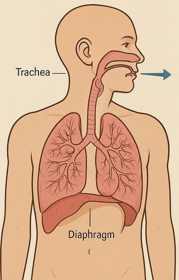
Air starts its path at the nose where we find its nasal cavity. Though one can breathe through the mouth, the nasal cavity is the main path, as there are vital protective and conditioning functions that the nasal cavity does.
As air moves to the nasal cavity, it moves through small openings that are lined using hair-like projections known as cilia and sticky mucus secreted by special epithelial cells. These structures combine to entrap dust, microorganisms, and other particles that would otherwise destroy the lungs in case of deeper inhalation in the respiratory tract.
The other significant role of the nasal cavity is air conditioning. The inner surfaces are lined with an intensive net-work of blood vessels which heat the incoming air to a approach to the body temperature. However, the mucus lining is also adding moisture which moisturizes the air and moves on to the deeper part of the lungs. This humidification allows the mucous membranes of the lower respiratory tract not to dry up.
Moreover, the nasal cavity has the sense of smell via an olfactory receptor in the upper part of the cavity. Although this sensory process does not play a direct role in the respiration, it may give advance warnings of dangerous substances in the air like smoke or chemicals
After being filtered, warmed and humidified, air enters the pharynx and the lower respiratory tract.
The Trachea: The Great Airway Canal
Once out of the pharynx, the air flows through the larynx and into the trachea commonly known as the wind pipe. The trachea is a primary airway that links the upper respiratory tract with the lungs.
C-shaped cartilage rings support the trachea in its structure. The presence of these rings ensures that the airway does not collapse during breathing and still experiences the freedom of movement as the neck swings. The anterior portion of every cartilage ring is opposite the esophagus, and the food rushes down the alimentary canal.
Trachea inner lining is made up of ciliated epithelial cells and goblet cells which produce the mucus. This is a combination that constitutes a defense mechanism commonly referred to as the mucociliary escalator. The dust and microorganisms that are absorbed in mucus are slowly pushed upward by cilia to the throat where they may be either swallowed or thrown out by coughing.
In this process, the trachea is significant in the further process of filtration that started in the nasal cavity. By the time the air has gone through this structure most of the large airborne particles have already been cleared.
The airway separates into two at the bottom of the trachea which are referred to as primary bronchi.
The Bronchi: Breathing into Every Lung
The trachea divides to form the right and right bronchi that direct to the right and left lungs respectively. These bronchi are seen to be major air distribution channels that take air further into the respiratory system.
The right bronchus is a little wider, shorter and more vertical than the left one. Due to such orientation, an inhaled foreign object has a higher possibility of getting into the right lung when accidentally aspirated.
The bronchi divide further into smaller structures within the lungs into secondary and tertiary bronchi. This parting trend is similar to an inverted tree and is also known as the bronchial tree.
The bronchi are also organized in the same manner as the trachea, the cartilage is supported, and the lining is covered with cells with cilia. But the closer the bronchi get the smaller the passages and the cartilage is less pronounced and the smooth muscle becomes a bigger contribution in regulating the airway diameter
This is a bifurcating network that gives even distribution of air in the lungs to enable all areas to be involved in the exchange of gases.
Bronchioles: Small Airways of the Lungs
The further division of the bronchi finally results in the creation of very small tubes known as bronchioles. These ducts measure less than one millimeter in diameter, and are the last conducting airways that occur prior to the gas exchange region.
As compared to the bigger bronchi, the bronchioles have no cartilage. They consist rather mainly of smooth muscle and elastic fibers in their walls. This structure enables the bronchioles to control the airflow by tightening or loosening.
As an illustration, when exercising the bronchioles open and permit more air to move through the lungs. Contrastingly, other conditions like asthma cause these airways to become narrower limiting the flow of air and thus making it hard to breathe.
Cilia and mucus-producing cells remain in the lining of bronchioles where, however, they decrease as the airways move towards the gas exchange areas.
Finally, the bronchioles terminate in minute air tubes called alveoli.
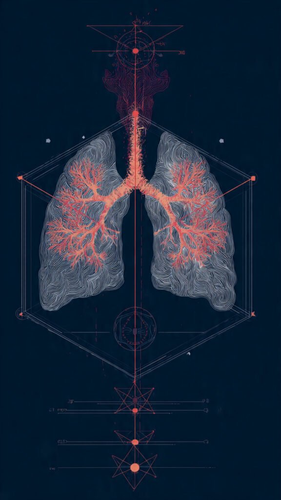
The Alveoli: The Gas Exchange Location
Pairs of tiny air sacs known as alveoli can be found at the end of the bronchioles. These structures are the main places of entry of oxygen into the bloodstream and elimination of carbon dioxide in the bloodstream.
exchange of gas in the alveoli and the contraction of the diaphragm during human breathing” width.
With in each lung being hundreds of millions of alveoli, it forms a huge surface area to carry out the gas exchange process, which is approximated to be about the size of a tennis court.
The alveoli have extremely thin walls, which are comprised of single layer of epithelial cells and a thick network of capillary network. Gases can easily pass across this thin barrier between the air contained in the alveoli and the blood contained in the capillaries.
The diffusion of oxygen into the blood takes place in the alveoli as oxygen concentration is more in the inhaled air than in the blood. Meanwhile, carbon dioxide is being carried out of the blood into the alveoli that will be exhaled later.
There are special cells in the alveoli which produce surfactant which is a substance that lowers the surface tension and prevents the air sacs that collapse during exhalation. Breathing without surfactant would have been much more difficult and the lungs would not be able to keep their air sacs open.
The alveoli facilitate efficient exchange of gases through this fine structure and huge surface area.
Ventilation and Pressure Gradients
The breathing process relies on the alteration of the pressure in the thoracic cavity. The air naturally moves to the high pressure zones to the low pressure zones. This principle is used by the respiratory system to move in and out air in the lungs.
Air enters the lungs when the pressures in the lungs are less than in the atmosphere. Air escapes when pressure on the inside is increased.
The differences in pressures are formed as a result of the alteration in the volume of the thoracic cavity, which can be mainly caused by the muscle activity.
The diaphragm refers to the most vital muscle in this process.
The Diaphragm in the Process of Breathing
Contraction of the diaphragm during the inhalation and exhalation movements.
The diaphragm is a massive dome shaped muscle that forms a separation between the thoracic and the abdominal cavity. It is used as the core of the ventilatory system and does the bulk of the air trade that takes place when breathing normally.
The muscle flattens downwards and moves during contraction of the diaphragm during inhalation. This motion raises the amount of the thoracic cavity and decreases the pressure within the lungs. Consequently, air is sucked into the respiratory tract.
In the exhalation process, the diaphragm relaxes and goes back to its dome-shaped position. This narrows the amount of air in the thoracic cavity making the internal pressure more intense and forcing the air out of the lungs.
This is done unconsciously and rhythmically in quiet breathing. In other cases like when a person is exercising, other muscles like the intercostal muscles between the ribs will help in the expansion and compression of the chest cavity.
A more in-depth expression of the structure and function of the diaphragm is available in this source on.
Breathing Process Co-ordination
Breathing mechanics are based on the accurate coordination of a number of components:
- Airway systems that move and refine air.
- Change in thoracic volume muscles.
- Driving airflows, pressure gradients.
- Alveoli which facilitate gas exchange.
Throughout inhalation, the contraction of the diaphragm causes the expansion of the thorax and the decreasing of the lung pressure that draws air to the lungs. The air passes through the nasal cavity, trachea, bronchi, and bronchioles where it is filtered, warmed and humidified.
When the air gets to the alveoli, oxygen is diffused into the blood stream whereas carbon dioxide is diffused out. The circulatory system then carries the oxygen rich blood to body tissues.
This process is reversed during exhalation and we lose carbon dioxide which is waste product of cell metabolism.
This recycling process repeats thousands of times a day, which makes sure that the oxygen that is required to cause energy is delivered to every cell.
The importance of Efficient Breathin
The breathing system should be able to sustain a fine balance. Oxygen supply to tissues might be impaired in case there is an obstruction of airflow, damaged alveoli, or the inability of the diaphragm to operate.
Asthma, chronic obstructive pulmonary disease (COPD), pneumonia, and respiratory infections are some of the conditions that disrupt one or more of the stages of the breathing process. There are diseases that constrict the bronchioles and others that destroy the alveoli or break or hamper the muscles which are used in ventilation.
Since the body uses oxygen to produce energy, any minor interferences with the breathing process can have extensive consequences on the general health.
The study of breathing mechanics, therefore, offers a great insight into the way the body sustains life, as well as the importance of ensuring a healthy respiratory system.
Conclusion
The breathing process is a complicated yet beautifully orchestrated physiological event that enables the body to get rid of gases in the environment and in the process acquire other gases. This air passes through the nasal cavity where it is filtered, warmed and humidified then moves along the trachea into the branching bronchi and finally into the lungs. There it passes on through bronchioles to alveoli which are openings in the lungs where oxygen is added to the bloodstream and carbon dioxide is eliminated.
Several gradients of pressures developed in the thoracic cavity cause the movement of air in and out of those structures. The changes in pressure do so to a significant extent because of the contraction of the diaphragm that is able to expand the chest cavity and consequently able to recoil during the exhalation.
The respiratory system is important in ensuring that oxygen gets to all the body cells through the joint effects caused by airway filtration, air conditioning, and the efficient exchange of gases in the body. In as much as the process is automatic, it is one of the most vital and the very delicate systems of human physiology- a process that keeps one alive with each breath.