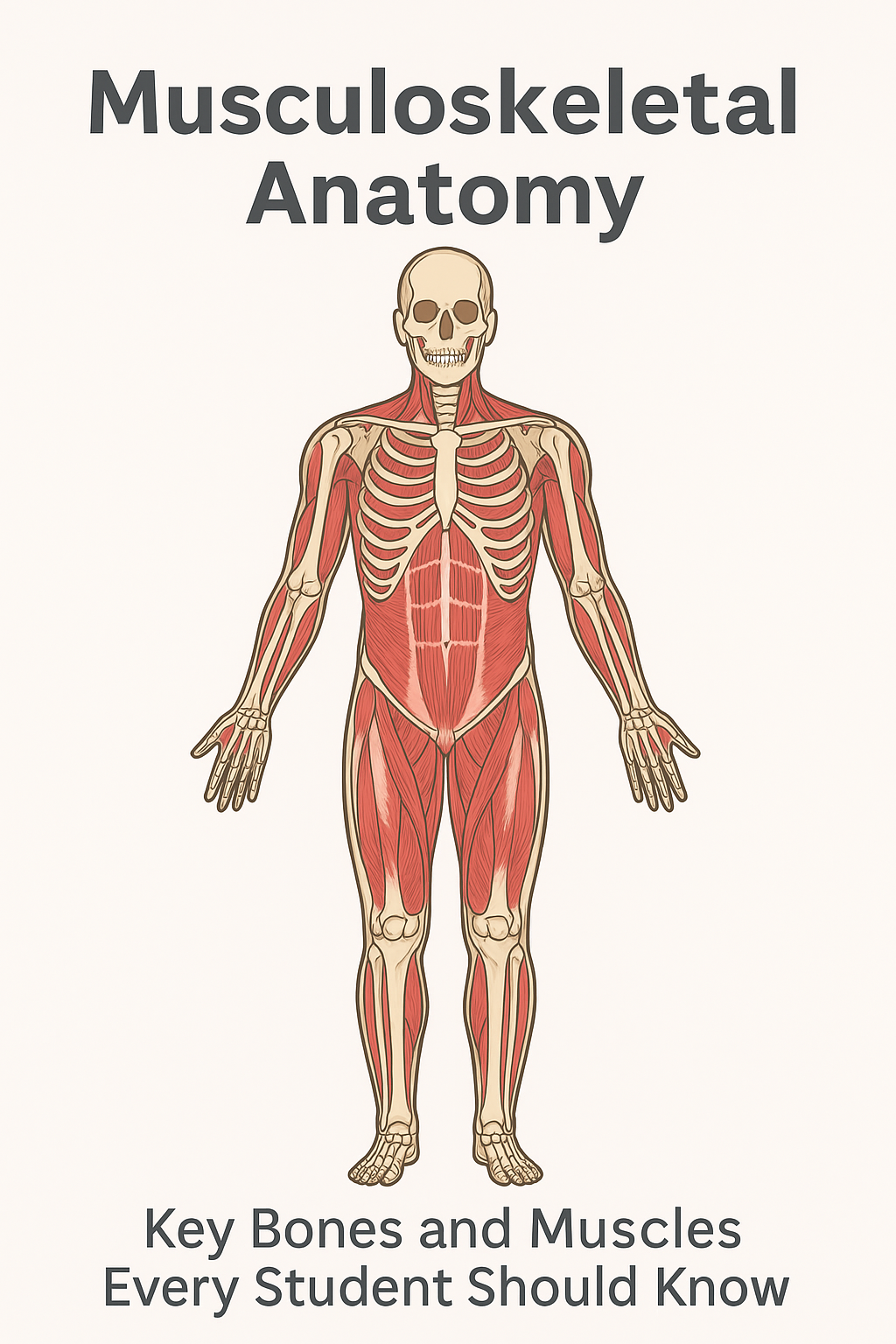
Knowing of the bones and muscles that are found in the human body is important to students learning biology, anatomy, physiology or planning to undergo clinical tests. Musculoskeletal system provides the mechanical basis of posture, stability, and movement and also safeguards the essential organs and anchors soft tissues.
The guide offers an easy yet high-paying dissection of the most significant bones and muscles, with an emphasis on the names and locations of the bones and muscles, as well as their functions – the very subjects that are regularly examined in medical and health-science programs.
In order to study more underpinnings, you can have a look at the following external source as well:
The following is the anchor link requested:
Introduction to the Musculoskeletal System
The musculoskeletal system entails:
- Bones – are hard structures that carry the body and protect internal organs.
- Muscles – these are tissues that are contracted to cause movement.
- Joints – these are the linkages between bones that enable movement.
- Tendons and Ligaments – connective tissues which stabilize the skeleton and move skeletal movements.
These structures work as a system which enables you to walk, lift objects, breathe, run and maintain balance.
SECTION 1: Bones that every student needs to know
Bones are not merely a kind of structure. These are muscular leverages, mineral deposits, and bone marrow. These are the bones that you should concentrate on in exams that have the highest yields.
Skull (Cranium)
Position: Head
Function: Safety of the brain, serves the organs of senses (eyes, nose, ears), composes the face.
Key bones to remember:
- Frontal bone – forehead
- Parietal bones – the sides and the top of the head.
- Temporal bones – ear surrounding areas.
- Occipital bone – the posterior part of the skull which has the foramen magnum.
- Mandible – lower jaw, which is the only mobile head bone.
Vertebral Column
Position: Midline of the back
Function: The spinal cord is protected, the body weight is supported, bending and rotation is possible.
There are 33 vertebrae in the vertebral column which are arranged as:
- Cervical (7) – neck
- Thoracic(12)- chest area, fixed to ribs.
- Lumbar (5) – buttock, holds the majority of the weight.
- Sacrum (5 fused)- forms the posterior portion of the pelvis.
- Coccyx (4 fused) – tailbone
High-yield concept:
The intervertebral discs are shock absorbers and they also provide flexibility.
Thorax (Chest Cage)
The thorax houses some of the essential organs such as the heart and lungs.
Key bones include:
- Sternum – breastbone
- Ribs (24 total)
- True ribs: 1-7
- False ribs: 8-10
- Floating ribs: 11-12
Rib movement is important in the functioning of the body during breathing.
Upper Limb Bones
The anatomy of the upper limb is very essential as the bones are what give fine motor control.
Shoulder Girdle
- Clavicle – collarbone
- Scapula – shoulder blade
Role: The arm should be stabilized and muscles should have a point of attachment.
Arm and Forearm
- Humerus – upper arm bone
- Radius and Ulna – bones in the forearms.
- Radius: thumb-side
- Ulna: pinky-side
Hand
- Carpals – 8 wrist bones
- Metacarpals – 5 palm bones
- Phalanges 3 fingers (2 on thumb), 2 on fingers.
Pelvis
Position: Lower trunk
Role: Provides stability to the spine and prevents pelvic organ damage and secures muscles to the lower limbs.
Key structures:
- Ilium – upper flared portion
- Ischium – the “sit bone”
- Pubis –ventral part forming pubic symphysis.
The childbirth mechanics cause differences in shape of the pelvis between biological males and females.
Lower Limb Bones
Thigh
- Femur – the longest and the strongest bone of the human figure.
Leg
- Tibia – shinbone
- Fibula – is a thin bone that offers lateral stability.
Knee
- Patella – kneecap, enhances force of the quadriceps muscle.
Foot
- Tarsals – ankle bones
- Metatarsals – midfoot bones
- Phalanges – toes
All these bones make it possible to bear weight, walk and maintain balance.
SECTION 2: The Important Muscles that every student must know.
There are several types of muscle, skeletal, smooth and cardiac but in the case of musculoskeletal anatomy we are talking of skeletal muscle, or those muscles that are attached to bone and cause voluntary movement.
The following are the muscles with the biggest yield:
Muscles of the Head and Neck
Sternocleidomastoid (SCM)
Position: Side of the neck
Function: Rotates and is able to flex the head.
Trapezius
Location: This is located at the back of the neck and high back of the back.
Action: Shoulders are raised, the scalpula moves.
Masseter
Position: Jaw
Location: This muscle is located in the mouth.
These are the muscles that are commonly found in face-to-face anatomy and physiology examinations.
Thoracic and Abdominal muscles.
Pectoralis Major
Position: Chest
Action: The arm is pulled inwards and across the body.
Intercostal Muscles
Position: Between the ribs
Function: Assistance breathing: Moving of ribcage.
Rectus Abdominis
Position: “Six-pack” muscle
Action: Flexion of the trunk; core stabilization.
Extrinsic and intrinsic Obliques
Location: Abdominal sides.
Function: Tilting and rotation of the trunk laterally.
Muscles needed are core muscles to maintain posture and avoid back injuries.
Muscles of the Upper Limb
Deltoid
Position: Shoulder
Function: Abducts the arm
Biceps Brachii
Position: Front of upper arm
Function Folds the arm and turns the forearm over.
Triceps Brachii
Position: Back of upper arm
Function: Extends elbow
Extensors and Flexors of Forearms.
Position: Forearm
Function: This is movement of the wrist, hand and fingers.
These are the muscles that are essential in performing duties that need accuracy such as writing or typing.
Muscles of the Back
Latissimus Dorsi
Position: Mid-lower back
Function: Pulls and stretches the arm (is used in swimming and climbing)
Erector Spinae
Location: The location is parallel to the spine.
Use: This is used to keep the back straight and maintain posture.
The back muscles are often tested due to their contribution to the posture and movement.
Lower Limb and Pelvis Muscles
Gluteus Maximus
Position: Buttocks
Action: Strausses and turns hip.
Quadriceps Femoris
Position: Front of thigh
Action: Exemplary knee extensors.
Components:
- Rectus femoris
- Vastus lateralis
- Vastus intermedius
- Vastus medialis
Hamstrings
Position: Back of thigh
Indication: Knee Flexion and hip extension.
Components:
- Biceps femoris
- Semitendinosus
- Semimembranosus
Gastrocnemius and Soleus
Position: Calf
Function: Plantarflexion (pointing the toes)
Tibialis Anterior
Position: Front of shin
Function Lifts the foot during ambulation.
These muscles are very much essential in walking, running, jumping and keeping balance.
Section 3: Bones and Muscles are functionally related
Musculoskeletal system operates by the mechanism of lever:
- Bones act as levers
- Joints act as pivots
- Muscles provide force
Examples:
- The contraction of the biceps brachii causes the pulling of the radius flexing the elbow.
- The quadriceps is an extensor of the leg, pulling the patella and tibia with the help of the patellar tendon.
- The heel is lifted by the gastrocnemius which acts on the calcaneus (heel bone).
Biomechanics, orthopedics and sports physiology are based on this relationship.
SECTION 4: Study tips to be used by students that are of high yield
Use Anatomical Landmarks
Memorize bony bumps like:
- Acromion
- Greater trochanter
- Medial malleolus
These show the essential muscle attachments.
Understand Movement Terms
- Flexion vs. extension
- Abduction vs. adduction
- Internal/ external rotation.
Such movements always have certain bones and muscles involved.
Practice With Diagrams
- Visual memory enhances the memory of the bone positions and the muscle orientations.
Connection Structure Clinical Relevance
Examples:
- Fractured radius has an impact on the rotation of the wrist.
- Strain in the lower back may be caused by tight hamstrings.
- Hip drop may be due to weak gluteus medius.
Knowledge of function simplifies and makes memory more significant.
Conclusion
Bones and muscles of the human body are a complex system supporting the posture, making it moving, and preserving vital organs. In case of exams of anatomy, medicine, physiotherapy, or biology, the best ground is that given to high-yield bones (such as the femur, the humerus, the skull, and the pelvis) and to major muscles (such as the deltoids, the quadriceps, the hamstrings, and the abdominals).
This article is presented in a concise, simplified, and exam-finding report that is supposed to guide you to study smarter and learn how skeletal structures and muscular groups collaborate with each other.