Movement is one of the defining characteristics of living. From the most basic action of blinking to the most complex action of performing athletic feats, every movement of the human body relies on the intricate coordination of muscle fibers, neural messages and biochemical processes. To the casual observer, muscle activity may look like a simple flex or extension but within each contraction is a sophisticated interplay of energy conversion, ion exchange and cellular machinery.
The science behind the process of muscle contraction, the methods the muscles use to contract, the different types of muscle fiber, and the effects of training on a physiological level give you the building block for a higher level of performance, injury prevention, and the true science behind the love of fitness. This article examines the fundamental mechanisms behind movement and how our training, recovery and nutrition impact muscular efficiency and endurance.
The Foundation of Muscle Function
The human body has more than 600 muscles that make up 40% of the total body weight. Each muscle is made up of bundles of fibers which can shorten upon stimulation by nerve impulses. This contraction, or shortening, produces force which produces movement. Muscle activity not only makes locomotion possible, but also helps keep the body in a stable posture, stabilize joint and regulate body temperature by heat generation.
The process that governs this function is called muscle physiology, the study of the process of muscles producing force and movement. It entails the knowledge of the structural organization of the muscle tissue, the biochemical reactions responsible for muscle contractions, and the adaptive responses to physical training.
Types of Muscle Tissue
There are three major types of muscle tissue in the human body: each of them has different characteristics and functions:
- Skeletal Muscle
Skeletal muscles are the voluntary muscles that are under the control of the somatic nervous system. They are connected to bones through tendons and they deal with movement and posture. These muscles appear striated when viewed under the microscope because of the periodic arrangement of contractile proteins – actin and myosin.
Skeletal muscles are under voluntary control and are capable of performing high-speed contractions with great force. Examples are the biceps muscle, the quadriceps muscle, and the pectoral muscles.
- Cardiac Muscle
Cardiac muscle is found in the walls of the heart. It is involuntary, striated, and has special intercalated discs that makes it possible to have synchronized contractions. These connections provide rhythm and efficiency to the heart in order to keep blood flowing throughout the body.
- Smooth Muscle
Smooth muscle is present in the walls of hollow organs such as the intestines, blood vessels and the bladder. It is without striations and involuntary. Smooth muscles are slow and sustained and are responsible for maintaining the functions of the body such as digestion, blood circulatory flow and respiration.
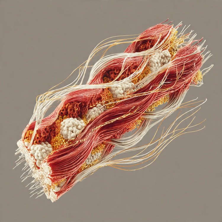
The Microscopic Anatomy of Skeletal Muscle
At the microscopic level, the skeletal muscle fibers are long cylindrical cells with multiple nuclei. Each fiber itself is composed of smaller units called myofibrils and inside myofibrils are contractile proteins actin (thin filaments) and myosin (thick filaments). These proteins are arranged in repeating units called sarcomeres, the basic contractile units of the muscle.
Actin and myosin filaments, which are the fundamental components of a sarcomere, overlap in a pattern of light and dark bands creating the striated appearance of the sarcomere seen in a microscope. When a muscle is contracting, these filaments move past each other – a process which has been termed the sliding filament theory.
The Process of Muscle Contraction
Muscle contraction phenomenon comprises of electrical, chemical and mechanical processes that collaborate in harmony. It happens in the following stages:
- Neutral Activation
The contractions of the muscle start with an electrical signal or action potential being dispatched by a motor neuron to the muscle, either by the brain or the spinal cord. The nerve conduction causes the signal to arrive at the neuromuscular junction; the point of nerve and muscle fiber intersection.
- Release of Neurotransmitters
It is at this intersection that acetylcholine (ACh) neurotransmitter is released into the synaptic cleft. ACh connects to the receptors of the muscle membrane and causes the occurrence of another electrical impulse that propagates along the muscle fiber.
- Calcium Release
The impulse is transmitted along T-tubules of the muscle and causes the sarcoplasmic reticulum, which contains calcium ions. Calcium is released to the cytoplasm, a choke of events is triggered leading to contraction.
- Cross Bridge Formation
Calcium binds to a protein known as troponin leading to a structural change in tropomyosin that reveals binding sites on actin filaments. These sites are bound by myosin heads and bind together in a cross-bridge.
- Power Stroke
The myosin heads pivot using energy provided by adenosine triphosphate (ATP) pulling the actin filaments inwards. This diminishes the length of the sarcomere and produces force – the apparent contraction.
- Relaxation
The withdrawal of the nerve impulse results in active pumping of calcium back into the sarcoplasmic reticulum. The actin binding sites are reoccluded, cross-bridges are dissociated and the muscle fiber restores its original state.
All this takes place within milliseconds and therefore muscle movement can be controlled very fast and accurately.
The Role of ATP in Muscle Function
The energy of muscle contraction is stored in form of ATP (adenosine triphosphate). In the absence of ATP, myosin heads would be bound to actin, which would cause rigidity a condition experienced following death, referred to as rigor mortis. The muscles constantly produce ATP in a variety of metabolic ways:
- Phosphagen System: implicates creatine phosphate utilized as a future energy source in brief and intense exercise (not exceeding 10 seconds).
- Anaerobic Glycolysis: Metabolism of glucose that involves the absence of oxygen to form ATP in a short period of time but produces lactic acid as a byproduct.
- Aerobic Respiration: is the use of oxygen in the mitochondria to generate ATP efficiently to be utilized during long time activities.
The endurance training will increase aerobic capacity whereas the strength training will increase the efficiency of the phosphagen system.
Types of Muscle Fibers
There are various types of fibers combined in skeletal muscles to define performance properties:
- Type I (Slow Twitch Fibers)
These fibers are very fatigue resistant and contract at a slow rate. They are mostly aerobic in nature and have a large number of mitochondria, myoglobin and capillaries. The endurance athletes like the marathon runners have type I fibers.
- Type IIa (Fast Twitch Oxidative Fibers)
These fibers are intermediate and they consist of both speed and moderate fatigue resistance. They are able to work aerobically and anaerobically hence they are good in activities that need power and stamina.
- Type IIb (Fast Twitch Glycolytic Fibers)
These fibers are fast contracting and produce high force although they wear out easily. Their primary metabolic process is anaerobic and are predominant in sprinters, weightlifters and power athletes.
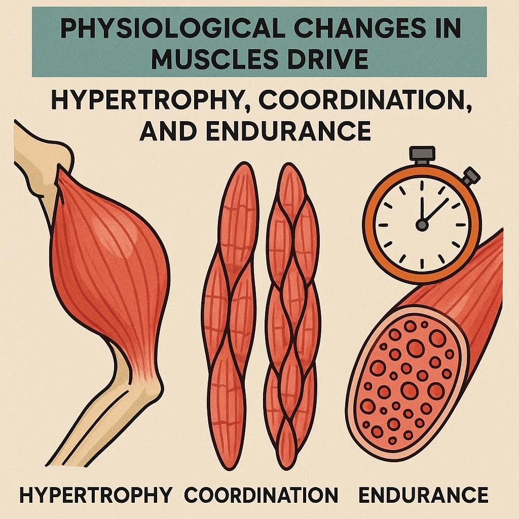
Adaptions to Training
The physical training also causes profound changes in the physiology of the muscles with improvement of power, stamina, and coordination.
Strength Training
Resistance exercises make muscles bulkier by hypertrophy or enlargement of the already existing muscle fibers. This is due to an augmented protein production and deposition of myofibrils. Neural activation is also enhanced by strength training that enables coordination and force production to be enhanced.
Endurance Training
The aerobic exercise improves the oxidative potential of the muscle fibres through augmenting mitochondrial density, capillary provision, and enzyme action. This helps the muscles to maintain long periods of physical activity and postpone fatigue.
Flexibility and Mobility Training
Stretching keeps the muscles and the joints in optimum position. It decreases the risk of strains, improves the posture and increases muscle contractions.
The Importance of Recovery
Training is important as training is recovery. Microscopic damage of muscles, which initiates repair and growth, occurs after vigorous exercise. This is enabled by an adequate rest, hydration, and nutrition. Sleep, especially, helps release of hormones (growth hormone) that helps in tissue repair and development.
On the other hand, overtraining causes fatigue, reduction in performance, and high risk of injury. Organized recovery rest: active rest, massage, stretching, etc. guarantees long-term improvement without health deterioration.
Injury Prevention and Muscle Maintenance
Balance the strengths and flexibility of healthy muscles require: training/rest, load/rest. The prevention of muscle trauma presupposes knowledge of physiological and mechanical aspects of movements.
- Warm Up and Cool Down
Proper warm-up raises the blood circulation, body temperature, and flexibility in muscles, making the body ready to be used physically. Resting the heart rate also aids in preventing stiffness that is caused by cool down.
- Balanced Training
Muscular imbalances might be caused by excessive attention to a specific form of exercise. Combining the strength and mobility exercises is a way of making sure that growth is even.
- Proper Nutrition
Protein consumption helps in repairing the muscles and carbohydrates restore glycogen storage. The nerve and muscle functions are maintained by electrolytes including calcium, potassium and magnesium.
Hormonal Influence on Muscle Growth
The role of hormones in repair and development of the muscles is crucial. The protein synthesis and tissue regeneration are stimulated by testosterone, growth hormone, and insulin-like growth factor (IGF-1). The stress hormone (cortisol) may impede muscle growth when high in chronic form, and balanced training and rest are crucial in this regard.
Aging and Muscle Physiology
Muscle mass and strength become natural as an individual grows old, a situation called sarcopenia. The cause of this process is hormonal, decreased activity and reduced protein synthesis. Nevertheless, frequent resistance and aerobic exercise can considerably reverse this loss, which maintains functional mobility and autonomy in the elderly population.
The Science Behind Stretching and Flexibility
Stretching enhances muscle elasticity and range of movement of a joint. There are two common types which are practiced:
- Static Stretching: Maintaining a stretch pose in order to stretch muscles and tendons.
- Dynamic Stretching: These are controlled movements which prepare the muscles to be active.
Stretching enhances blood flow and decreases stiffness as well as improves proprioception- body awareness of position and movement. Stretching is the best way to performance and injury prevention, which is best done in the warm-up and the cool-down.
Integrating Mind and Muscle: The Neuromuscular Connection
All the voluntary muscle movement starts on the side of the brain. Motor neurons send signals to muscle fibers and adjust the timing and muscle strength. This neuromuscular connection increases with the frequency of the movement. This concept is called muscle memory which forms the basis of athletic skill and recovery of athletic ability after injury.
This connection can be further improved with the help of visualization and mindfulness techniques, which will increase the performance and level of accuracy during the movements.
The Role of Hydration and Electrolytes
Muscle activities cannot work without water and electrolytes. The changes in the electrolytes may cause cramps or weakness, and the dehydration causes the decrease of blood volume and the impediment of oxygen delivery. Hydration helps in metabolism of energy and control of temperature in the course of exercise.
Conclusion
In the field of science of muscular physiology, movement is much more than mere mechanical movement, it is a symphony of electrical messages, chemical energy and biological adaptation. Every contraction manifests the exceptional ability of the body to coordinate and be resilient.
Training makes this system ever more challenging; recovery makes it ever more refined by growing and repairing. In any case, be it using strength training, endurance exercises, or conscious stretching, by learning the mechanisms of muscle action one can exercise effectively, avoid injuries, and maintain mobility throughout a lifetime.
With the balance between effort and rest, we can admire the amazing design of the body the design which enables us to move, create and live by breathing every breath and by walking every step.