Introduction
Periodontal disease is a persistent inflammatory disease that involves the supporting of the teeth as the gingiva (gums), periodontal ligament and bone of the alveolar. It starts as gingivitis characterized by inflammation of the gum and may develop further to periodontitis where permanent destruction of the tissue may take place and may lead to loss of teeth. Way beyond the oral issue periodontal disease has now been identified as a systemic health problem that is connected to cardiovascular diseases, diabetes, and poor pregnancy outcomes.
The periodontal disease development and progression is primarily influenced by a complicated combination of pathogenic bacterial biofilms and the immune-inflammatory response of the host. It would thus be necessary to find out the molecular pathways of periodontal disease in order to formulate more effective diagnostic biomarkers and therapeutic interventions.
The Bacterial Biofilm and Periodontal Disease
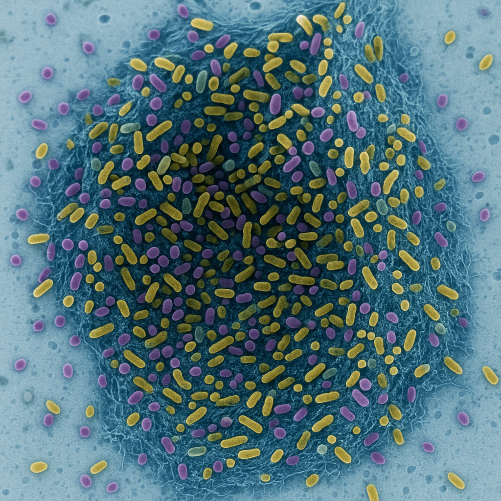
Periodontal disease begins with the development of bacterial biofilms which are also referred to as dental plaque on the tooth surface. These biofilms are complex microbial communities that are surrounded by a self produced extracellular matrix of polysaccharides, proteins, and nucleic acids. Biofilm-associated microbes have a higher resistance to antibiotics and the immune defense of the host, unlike the free-floating bacteria.
Examples of important periodontal pathogens are:
- Porphyromonas gingivalis
- Aggregatibacter actinomycetemcomitans
- Tannerella forsythia
- Treponema denticola
The virulence factors that are discharged by these pathogens include lipopolysaccharides (LPS), proteases and fimbriae which stimulate the host cells especially the gingival epithelial cells and immune cells. An example is LPS that interacts with Toll-like receptors (TLRs) on host cells, which cause the release of inflammatory mediators.
Besides, biofilm formation modifies the microenvironment of the gingival sulcus, which results in a reduction of oxygen and an increase in the growth of anaerobic bacteria. The constant presence of these bacteria provokes the chronic immune response, and this leads to the formation of the chain reaction of inflammation and tissue destruction.
Bacterial Biofilm and Periodontal Disease
The first malefactor in periodontal disease is bacterial biofilms. They are well-organized communities of bacteria entrenched in a polymorphic extracellular polymer (EPS) that increases their antibiotic and immune resistance. The most frequently occurring pathogenic species are Porphyromonas gingivalis, Tannerella forsythia, and Treponema denticola- also known as the red complex.
Biofilm Formation and Virulence
Biofilm formation occurs in different bacterial species, and its presence differs according to the microbe, thereby affecting virulence. Biofilm Formation and Virulence Biofilm formation is a process that takes place in various bacterial species, and its occurrence varies with the microbe, thus influencing virulence.
Biofilm lifecycle is divided into phases- initial adhesion, colonization, maturation and dispersion. When formed these biofilms secrete virulence factors including:
- Lipopolysaccharides (LPS): Cause great inflammatory reaction.
- Proteases (e.g., gingipains): Breaks down the host proteins and immune factors.
- Short-chain fatty acids: Regulate the work of immune cells.
These molecules interfere with the barriers of the epithelial and activate the production of the pro-inflammatory cytokines, enhancing the destruction of the tissues.
Host Immune Response: The Two Sided Sword
An immune reaction to bacterial invasion is essential in avoiding infection but in periodontal disease, the hyperstimulated or hyper-regulated reaction causes tissue destruction.
2.1 Innate Immune Activation
Pattern recognition receptors (PRRs) TLR2 and TLR4 identify bacterial molecules like LPS and peptidoglycan when the bacterial biofilm components interact with the host cells. This identification triggers intracellular signaling pathways and more specifically the nuclear factor-kappa B (NF-kB) pathway which leads to the expression of pro-inflammatory genes.
The recruitment of activated neutrophils and macrophages to the gingival crevice occurs, and the cells are aimed at destroying bacteria by phagocytosis and reactive oxygen species (ROS) release. Nevertheless, high levels of ROS may cause harm to the host tissues, causing oxidative stress and additional inflammation.
2.2 Adaptive Immune Response
In the long run, it is followed by adaptive immunity. Dendritic cells and other antigen-presenting cells (APCs) process bacteria antigens in order to present them to T-cells which are differentiated into several subtypes:
- Th1 cells, which stimulate cell mediated immunity through interferon- gamma (IFN-g).
- Vigorous B-cell antibodies are triggered by Th2 cells.
- Th17 cells, which secrete interleukin-17 (IL-17), which augment the recruitment of neutrophil, but also induce tissue damage.
These subsets of T-cells lead to the prolonged release of pro-inflammatory cytokines, which perpetuates the destruction cycle.
The Important Inflammatory Mediators and Cytokines
Periodontal disease is characterized by inflammation, and a number of cytokines and inflammatory mediators have crucial roles in its pathogenesis.
3.1 Pro-inflammatory Cytokines
- Interleukin-1 beta (IL-1b): Mediates osteoclast activation, and results in bone resorption.
- Tumor necrosis factor-alpha (TNF-a): Makes it permeable to the vascular system and brings more immune cells into the area of infection.
- Interleukin-6 (IL-6): B-cell differentiation mediator and enhances the acute-phase protein synthesis in the liver.
High concentrations of these cytokines in the gingival crevicular fluid are a disease severity indicator and are the possible biomarkers of diagnosis.
MMPs play a role in breaking down food components and protein fibers related to the skin, bone, and cartilage (Bray, 2007).
3.2 Matrix Metalloproteinases (MMPs)
MMPs are involved in the breakdown of food and protein fibers in and around the skin, bone and cartilage (Bray, 2007).
The neutrophils and fibroblasts secret exudative enzymes known as MMPs that destroy the components of the extra cellular matrix like collagen. MMP-8 and MMP-9 play a significant role especially in the destruction of periodontal tissues. In the normal state, tissue inhibitors of metalloproteinases (TIMPs) regulate their activity but in pathological conditions, this process is disrupted leading to unregulated tissue breakdown.
3.3 Lipid Mediators
Prostaglandin E2 (PGE2), which is the product of cyclophosphamide of arachnoid acid, increases inflammation and bone resorption. PGE 2 levels are highly correlated with active periodontal lesions.
The Destruction of Tissues on a Molecular Level
Periodontal tissue destruction is a cascade of molecular pathways that are caused by both bacterial virulence factors and host immune responses.
4.1 NF-kB Signaling Pathway
NF-kB is a major control of inflammation. Upon triggering by bacterial elements such as LPS, NF-kB moves to the nucleus, and induces the expression of cytokine, chemokine, and adhesion molecule encoding genes. It causes sustained recruitment and intensification of inflammation in immune cells.
4.2 MAPK (Mitogen-Activated Protein Kinase) Pathway
It is activated by growth hormone-releasing hormone (GH-RH), a growth hormone secretagogue, and suppressed by insulin and reelin through inhibition of 4.2 MAPK (Mitogen-Activated Protein Kinase).
The MAPK signalling pathway (which incorporates ERK, JNK and p38 residues), transduces the cellular reaction to stress and cytokines. It enhances MMP expression, which leads to the connective tissue degradation.
4.3 Bone resorption RANK/RANKL/OPG Axis
Periodontitis bone resorption is controlled by RANK (Receptor Activator of Nuclear Factor Kappa-B) and its ligand RANKL that are expressed on osteoblasts and activated T-cells. RANKL interacts with RANK on osteoclastic precursors, and it promotes the development of bone-resorbing osteoclasts.
Osteoprotegerin (OPG) is a decoy receptor that attaches to RANKL, and this prevents RANKL from interacting with RANK. In periodontal disease, the disproportion in the ratio of RANKL and OPG contributes to increased bone resorption.
Oxidative Stress and Apoptosis
Reactive oxygen species (ROS) that are generated by activated neutrophils and macrophages have a dual role in periodontal disease. Though critical in bacterial killing, excessive ROS harms lipids, proteins and DNA of the host cells. This oxidative stress causes the activation of apoptotic pathways in gingival fibroblasts and osteoblasts of tissue repair processes.
Indicators malondialdehyde (MDA) and 8-hydroxydeoxyguanosine (8-OHdG) are higher in periodontal tissues, which are indicators of lipid peroxidation and oxidative damage of DNA respectively. The oxidative stress pathways are also coming under the scope of investigation as a form of therapy.
Epigenetic and Genetic Regulation
Recent studies have shown a role of epigenetic changes in periodontal disease – DNA methylation, histone acetylation, and microRNA up- or down-regulations have been observed to modify activity of inflammatory genes. For example:
- SIL-6 and TNF-a gene promoters are hypomethylated and their expression is up-regulated.
- TLR signaling and cytokine production is controlled by microRNAs, including miR-146a.
Genetic predisposition is also a factor as the polymorphism of genes of proinflammatory cytokines (e.g. IL-1b) is associated with high risk of developing severe periodontitis.
Potential Diagnostic Markers
The discovery of molecular biomarkers is very important in early diagnosis and tracking of disease progression. Examples of biomarkers in gingival crevicular fluid (GCF), saliva, or serum are:
- Cytokines (IL-1b, TNF-a, IL-6)
- MMPs (particularly MMP-8)
- Oxidative stress products (MDA, 8-OHdG)
- Bacterial DNA of most important pathogens.
Salivary diagnostics in particular is a developing non-invasive and economical technique of identifying early periodontal alteration prior to the development of clinical symptoms.
Augmenting Therapeutic Targets
The developments in molecular biology have provided avenues to more selective and efficient periodontal therapies than the conventional scaling and root planing that has always been associated with periodontal therapy.
Possible molecular-based treatments are:
- Cytokine-blockers: TNF-a or IL-1b-inhibitors to stop inflammation.
- To inhibit connective tissue degradation: MMP inhibitors.
- Antioxidants: These include resveratrol and coenzyme Q10 in order to fight oxidative stress.
- Host modulation therapy: Inflammatory mediators are modulated with the use of agents such as sub-antimicrobial dose doxycycline.
- Probiotics and biofilm disruptors To reestablish the microbiome of the oral cavity and avoid recolonization with the harmful species.
Additionally, gene therapy and epigenetic drug studies are underway to regulate the activity of pro-inflammatory genes, which can provide the opportunity to control the disease in the long-term.
Conclusion
Periodontal disease is much more than a basic bacterial infection, a multifactorial, immuno-inflammatory disease whose pathophysiology is controlled by the complex of molecular and cellular interactions. The interactions of the bacterial biofilms with the host immune responses, oxidative stress, and cytokines lead to progressive tissue destruction and bone loss.
Thorough knowledge of such molecular processes can not only clarify the pathogenesis of the disease but also gives a chance to diagnose early, provide individual treatment, and new therapeutic objects. With the ongoing research in deciphering the molecular basis of periodontal disease, clinicians are letting go of the goal of precision medicine that has the potential of sustaining oral and systemic health.