The profound knowledge of head and neck anatomy is one of the foundations of contemporary medicine. This area is very complex with structures that harbor vital organs, neurovascular systems as well as functional systems that support speech, swallowing, respiration and sensory sensations. To healthcare practitioners, especially those in the field of medicine and dental practice, understanding of such anatomical facts is necessary to avoid misdiagnosis, preventive treatment and surgical intervention.
This article discusses how the study of head and neck anatomy can be used in clinical practice, focusing on surgery, diagnostics, emergency care, and dental care.
Significance of Head and Neck Anatomy in the Medical Field
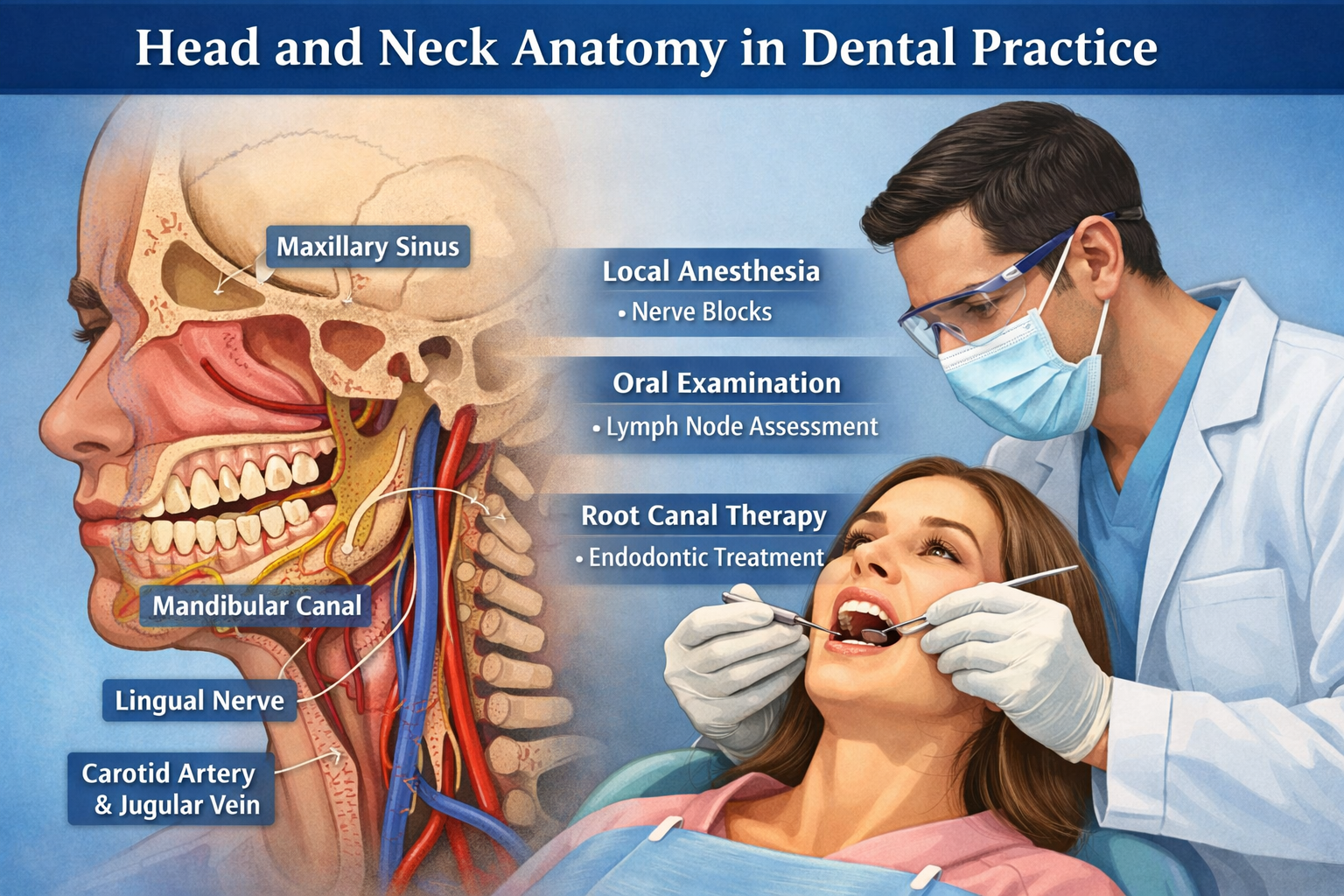
The cranial structures, the cervical spine, the great arteries and veins, the lymphatic system, the muscles, the nerves, and the sensory organs make up the head and neck region. All medical or dental operations in this region must be done with special attention to these elements in consideration of their density and functionality.
As an example, the facial nerve (cranial nerve VII) and trigeminal nerve (cranial nerve V) of cranial nerves provide sensation, mastication, and facial expression. Surgery on these nerves may lead to permanent deficits and this underscores the importance of anatomy as a source of clinical safety. In the same manner, the carotid arteries and jugular veins supply and drain major blood supply and hence, close anatomical orientation is critical to prevent disastrous complications.
Surgical Practice Applications
Oral Surgery and Maxillofacial
Surgery of the oral cavity, the jaw, and bones of the face require comprehensive knowledge of local anatomy. Treatment of fractures, corrective surgery on the jaw (orthognathic surgery), and tooth extraction are procedures based on the space relationship of teeth and alveolar bone, nerves, and blood vessels.
Poor alveolar nerve consideration during molar extraction in dental practice may result in loss of sensation in lower lip and chin. Equally, the maxillary sinus anatomy can be very essential during such operations as sinu lifts or when the implant is to be placed so as to avoid sinu perforation.
ENT and Neck Surgery
Head and neck surgeons handle complicated surgeries that include thyroid, parathyroid glands, larynx and cervical lymph nodes. The anatomical knowledge can be used to excise the tumors safely, deal with airway obstructions, and maintain functioning. In thyroidectomy, one can give an example of the recurrent laryngeal nerve as it is imperative to avoid paralysis of the vocal cords.
Moreover, the knowledge of the cervical fascial planes enables the surgeons to treat infections, drain abscesses, or even carry out a reconstructive surgery effectively. Such structures act as the avenues of contagion of infections or tumors and identification of these avenues can lead to both pre-operative surgical planning and after-surgical treatment.
Diagnostic Applications
Imaging Interpretation
Clinicians and radiologists use anatomy to interpret an imaging study like CT scan, MRI, and X-rays. E.g. to be able to distinguish between benign and malignant lesions in the neck, the anatomy of the salivary glands, the location of the lymph nodes and the normal anatomy of the cervical spine.
In the dental radiology field, proper understanding of panoramic and cone-beam CT scans enables the dental practitioner to examine the quality of the bone, locate the impacted teeth and also determine the closeness to the nerves and the sinuses. Diagnosis or incorrect treatment plan may result without specific anatomical knowledge.
Physical Examination and Palpation
Physical assessment is based on anatomical expertise. The lymph nodes, thyroid, and salivary glands palpation are used to identify infections, tumors, and structural abnormalities. Mediators who have been trained in anatomy are able to differentiate normal variations and pathological changes so that they can intervene on time.
To illustrate, in dental practice, the palpation of the submandibular gland or anterior cervical lymph nodes may help give an indication of infection or malignancy to aid more specific research.
Trauma management and Emergency Care
Traumatic head and neck injuries are frequent occurrences that result because of accidents, falls, or sporting events. In order to avoid secondary complications, emergency physicians have to move in this direction with precision.
Airway Management
A critical issue in trauma is the obstruction of airways. The anatomical understanding of the pharynx, the larynx, and the trachea enables medical practitioners to intubate safely, or to handle emergency surgical airways. Instruments may be misplaced and result in bleeding, aspiration and permanent injury to the vocal structures.
Hemorrhage Control
Extensive hemorrhage due to a laceration of the face or neck requires instant awareness of the vascular structure. The key skills of trauma management are the ability to have rapid control of the carotid or jugular vessels and the ability to identify the compressible zones and awareness of the underlying nerves.
Oral injuries that are minor can cause serious hemorrhage in case of injury to arterial branches, like facial artery. Dental practitioners should thus be ready to deal with such emergencies, more so in outpatient facilities.
Cervical Spine Protection
Trauma usually touches on cervical vertebrae that surround the spinal cord. Knowledge of the correlation between the cervical bones, ligaments and the neurovascular structures surrounding them facilitates immobilization and minimizes the probability of permanent paralysis. Such information is of especial interest to prehospital care providers and emergency room clinicians.
Dental Practice Applications
Local Anesthesia
Successful management of local anesthetic must have accurate understanding of the anatomy of the head and neck. Dental blocks like the inferior alveolar nerve block, posterior superior alveolar nerve block or infraorbital nerve block are based on accurate anatomical placement. The loss may lead to partial anesthesia or damage of nerves and vessels.
Periodontal and Prosthodontic Surgeries
The bone density, gum surgery, and nerve pathways must be known in cases like tooth extractions, gum surgeries and placing of prosthetic devices. As an example, the maxillary sinu anatomy can be used to avoid perforation and postoperative complications in sinu floor elevation prior to dental implant placement.
Oral Pathology Detection
The anatomy is needed in early detection of oral cancers or pre-malignant lesions. In order to detect suspicious areas, dentists need to check in mucosal surfaces, salivary glands apertures, and lymphatic drainages. Early detection of abnormal changes in tissues promotes timely referral to a specialist and patient outcomes.
Temporomandibular Joint (TMJ) Management
TMJ is a convoluted articulation and it must understand the structure of the bones, the mastication muscles and ligaments. This knowledge is applied by the dental professionals to diagnose the disorders, prepare the surgery, or apply the orthodontic treatment. Proper anatomical understanding can avoid joint injuries and can assist in functional recovery.
Control of Lymphatic System and Infection
The number of lymph nodes in the head and neck is very high and is arranged in the form of cervical chains. The structures are important in the management of infections and the staging of the cancer. As one instance, swollen lymph nodes in the neck may demonstrate general infections, cancerous lesions of the mouth or metastasis. Clinicians and dental practitioners should be in a position to identify between normal reactive nodes and pathological enlargement.
The knowledge of the lymphatic drainage patterns is also informative in surgical planning. Precisely, the surgeons are able to excise lymph nodes and do it without causing excessive tissue damage and they are able to do so with adequate oncologic control. This especially applies in oral and oropharyngeal cancers whereby cervical lymph nodes are likely to contain metastases.
Nerve Conservation and Rehabilitation
The head and neck anatomy is critical in avoiding nerve damage during clinical practice. Some of these nerves include the facial, glossopharyngeal and hypoglossal nerves, which govern some of the key activities that include facial movement, taste, swallowing, and speech.
Long term disability can be caused by accidental nerve damage. Anatomical variation knowledge enables surgeons and dentists to reduce the risks, repair nerve damage, and rehabilitate. Rehabilitation may involve speech therapy or physical therapy, and this may involve the knowledge of normal nerve and muscle structure to target the problem.
Interdisciplinary Relevance
Anatomy of the head and neck not only plays a significant role in the work of surgeons and dentists but also that of other medical workers, such as:
- Radiologists: To interpret the images correctly, it is dependent on anatomical landmarks.
- Speech and language therapists: The anatomy of the oral cavity, pharynx and larynx is applicable in the rehabilitation of speech and swallowing disorders.
- Oncologists Anatomical knowledge determines biopsy, staging, and radio-planning.
- Emergency responders: The management of injuries in the head and neck is based on the quick evaluation of the anatomy.
This interdisciplinary applicability ascertains the significance of sound anatomical education in medical and dental training.
Conclusion
Anatomical knowledge of the head and neck is invaluable in current healthcare. It supports safe operating practice, proper diagnosis, efficient emergency treatment and effective dental practice. In the case of dental practitioners, their understanding of the oral structure, nerves, and vascular networks has a direct effect on patient safety and success of treatment.
Likewise, surgeons, doctors and other health practitioners use anatomical knowledge in handling trauma management, cancer and functional rehabilitation. Never-ending learning, practical training, and application of anatomy to clinical practice will help to enable medical professionals to provide accurate, effective, and patient-centered services.
Finally, solid Head and neck anatomy is a pillar that helps in the transition between theory and patient excellence, protecting the patient outcomes and promoting the quality of healthcare delivery.