Scientifically, tooth development or odontogenesis is a complicated biological process that commences at an early stage of embryonic life into the childhood period when teeth would finally erupt. Learning the microscopic structure of teeth at different developmental stages is important to both clinicians and researchers since it enables the detection of developmental defects, including enamel hypoplasia, dentin dysplasia, and other inborn dental pathologies. This paper explores the phases of the development of the tooth with special focus on the histological processes that take place during the period between the initiation and eruption.
Development of Teeth: Basics
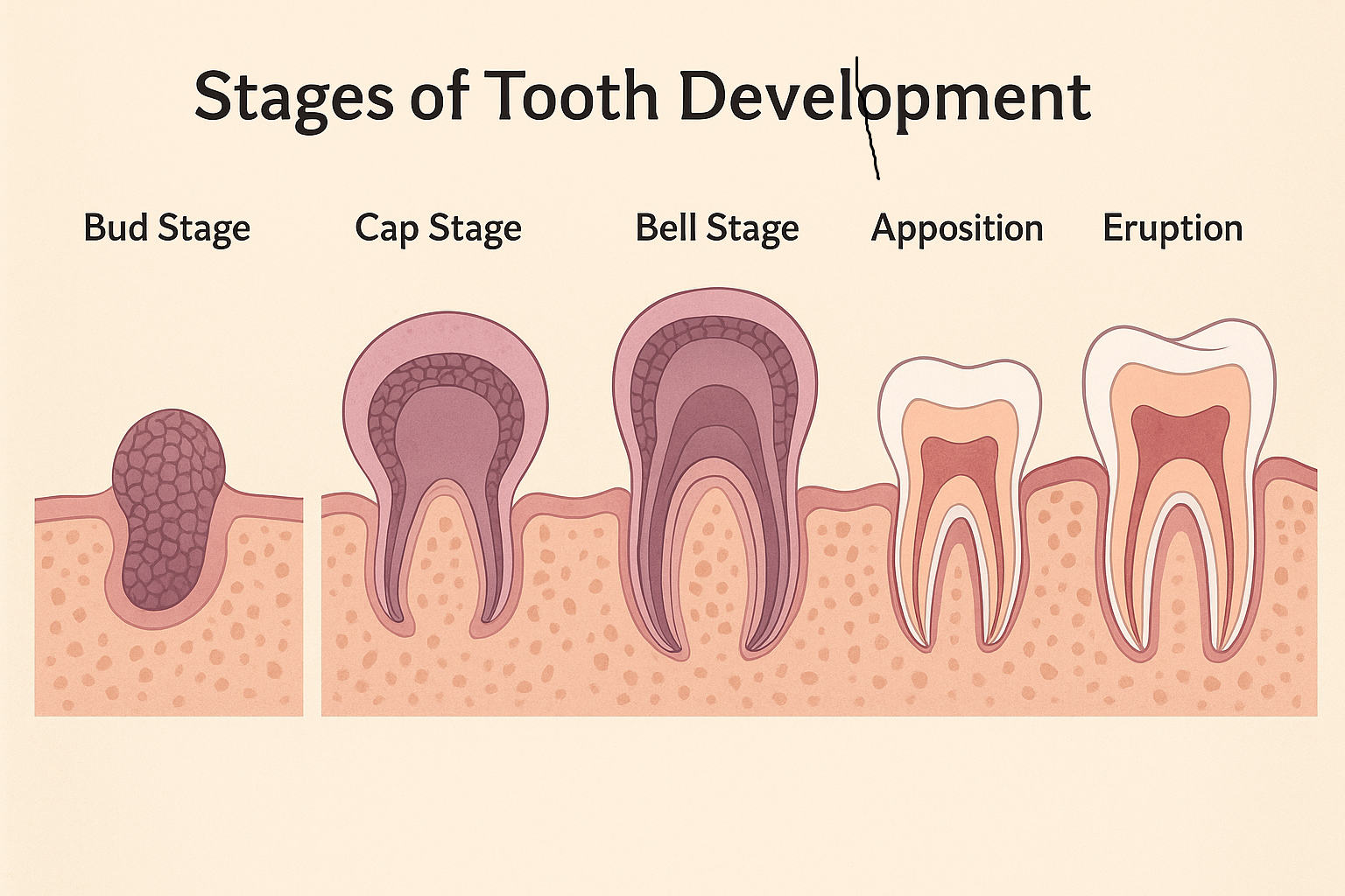
The formation of teeth consists of well-coordinated communications between the mesenchymal and epithelial tissues. These tissues communicate by means of molecular signaling pathways in order to have the dental structures formed properly. Histologically, the development of the odontogenesis could be differentiated into successive stages: bud, cap, bell, apposition and eruption. The different stages have their own specific cellular arrangements and patterns of tissue differentiation.
It is a good idea to get a basis of the stages of tooth development as compared to that of the embryology before getting into the histology of each stage. These steps do not only outline how morphological changes occur but also give understanding of the interactions that surround normal tooth formation in tissues.
Stage 1: The Bud Stage
The earliest stage that can be determined in tooth development is the bud stage which takes place at about the sixth week of embryonic life in primary teeth. This is where oral epithelium becomes thickened at this area to develop dental lamina, a tract of epithelial cells that invagy into the underlying mesenchyme.
Histological Features:
- Dental lamina is seen as an extension of basal epithelial cells into the mesenchyme.
- The epithelial bud is covered by mesenchymal cells, which are loose in structure and undifferentiated and subsequently become the dental papilla.
- At this point there is no development of enamel or dentin, but the emphasis is on epithelial-mesenchymal signaling.
Bud stage lays the groundwork of future shape and location of teeth. At this stage, interference may cause agenesis (absence of teeth) or supernumerary teeth and hence the significance of tissue-level regulation at the beginning of development.
Stage 2: The Cap Stage
The bud enters the cap stage in the ninth to tenth week of development which is so called because of the appearance of the cap-like structures of the epithelial cells that cover the dental papilla. This is the initiation point of morphodifferentiation in which the fate of the type of tooth begins to be established.
Histological Features:
The epithelial bud is then expanded to create the enamel organ, which then differentiates to give three different layers:
- Outer enamel epithelium (OEE): This is a layer of cuboidal cells which is protective in nature.
- Stellate reticulum: Star-shaped cells creating a loose and hydrated structure of network, which facilitates the formation of enamel.
- Inner enamel epithelium (IEE): Columnar cells which are to be differentiated into ameloblasts, the secretory cells of enamel.
- The dentin and pulp tissue will develop out of the condensed dental papilla beneath the enamel organ.
- The dental follicle is surrounded by mesenchymes and will have a role to play in periodontal ligament, cementum and alveolar bone.
- The cap stage is the histological evidence of the first cell specialization, which is essential in the formation of enamel and dentin. The abnormalities at this stage can result in dens in dente or malformed crowns.
Stage 3: The Bell Stage
The bell stage is the period between 14 th and 18 th weeks of development, which is further broken down into an early and late phase. This is the stage at which the tooth germ becomes bell-shaped and histodifferentiation and morphodifferentiation broaden.
Histological Features:
The cells of inner enamel epithelium (IEE) cells lengthen and develop into pre-ameloblasts, which develop into ameloblasts, and the production of the enamel matrix starts.
- The dental papilla divides into odontoblasts and they initiate the deposition of dentin.
- Stellate reticulum goes on to give enamel-forming cells cushioning and metabolic support.
- The outer enamel epithelium (OEE) is protective and the cervical loop, where OEE and IEE meet, is Hertwig epithelial root sheath, necessary in the formation of the root.
This phase is very crucial since it determines the final shape of the tooth crown and it is followed by a series of deposition of hard tissues. Histological changes in this case may lead to either amelogenesis imperfecta (deformed formation of enamel) or defects of dentin.
Stage 4: The Apposition Stage
The stage that comes after the bell stage is the apposition stage which entails the actual secretion of the enamel and dentin matrices. It is a period of severe cellular activity and tissue deposition.
Histological Features:
- The enamel matrix released by Ameloblasts is very well organized along the dentino-enamel junction (DEJ).
- Predentin is secreted by the odontoblasts and later on mineralized to give rise to dentin.
- The dental papilla is now changed to dental pulp which contains the fibroblasts, blood vessels and nerves.
- The tooth germ is surrounded by a dental follicle that is involved in the formation of cementum, periodontal ligament, and alveolar bone.
Histologically, the apposition stage demonstrates the interaction of the ameloblasts and odontoblasts which is necessary to normal development of the crown. Disruptions during this stage can result in the enamel hypoplasia which is a condition that leads to a thin or pitted enamel.
Stage 5: The Maturation Stage
Mineralization takes place in the maturation stage after the process of apposition of enamel and dentin. This stage is to make the hard tissues attain their end density and strength.
Histological Features:
- The ameloblasts also regulate to eliminate water and organic substances in the enamel and as a result hydroxyapatite crystals develop.
- Odontoblasts keep on with dentin mineralization and preserve the dentinal tubules.
- Dental pulp is another tissue which is still important to aid the growing tooth.
The inability to pass the maturation stage may result in hypomineralized enamel, which predisposes teeth to caries and mechanical damages. Histology at this point aids in diagnosing these defects on a cellular level.
Stage 6: Formation and Eruption of Roots
Once it gets formed, the focus shifts to the development of the roots and subsequent eruption of the teeth. Root shape is regulated by the Hertwig epithelial root sheath (HERS) which triggers odontoblast differentiation in the root area.
Histological Features:
- The deposition of root dentin is done by odontoblasts and the regression of ameloblasts occurs because the enamel is not formed extending into the root.
- Dental follicle is differentiated into periodontal ligament, cementum and alveolar bone to nourish the developing tooth.
- The process of eruption is complicated by bone resorption with the participation of the osteoclast and a cellular activity in the periodontal ligament.
History Abnormalities in the HERS or root-dentin formation may cause the root malformations, delayed root eruption, or impaction, which is why the processes of the tissue level are important in clinical outcomes.
Tooth Histology as Clinical Implications
Having knowledge of the stages of tooth formation in the histological context enables dental specialists to detect and treat developmental dental problems effectively. Key conditions include:
Enamel Hypoplasia
- Brought about by troubles during the apposition or maturation phases.
- There is decreased enamel thickness with abnormal prism formation which is seen under histology.
Dentin Dysplasia
- The outcome of odontoblast malfunctioning occurs at the bell stage or the apposition phase.
- The histology shows the abnormal pattern of dentinal tubules and anomaly of pulp chamber.
Congenital Tooth Agenesis or Supernumerary Teeth
- Grow as a result of the breakdown of bud or cap stages.
- Histologically, dental lamina may be missing or epithelial may be overgrown.
Amelogenesis Imperfecta
- The ameloblast failure is either in the apposition or maturation phases.
- Poorly formed enamel matrix or incomplete mineralization is found using histology.
Through a comparison of clinical observations and histological observations, the dentists may offer specific therapies and preventive measures.
Conclusion
Odontogenesis is a carefully planned series of histological processes through which undifferentiated, epithelial and mesenchymal cells are differentiated to give rise to fully functioning teeth. The different phases, such as the bud, eruption, etc., have particular cell organization, tissue differentiation, and molecular interactions. A disturbance at any stage can result in developmental aberration and hence the necessity of using histology to diagnose as well as in the planning and treatment of diseases.
The comprehensive knowledge of the tooth development stages does not only add to the academic knowledge but also has a direct influence on clinical practice. Histological understanding can be used by dentists and scientific researchers to estimate, diagnose, and control diseases like enamel hypoplasia, dentin dysplasia, and other inborn dental defects, which will lead to better oral health outcomes.