Odontogenesis, or the tooth formation, is one of the most complex developments in human biology. It is a timely interaction between epithelial and mesenchymal tissue, which is controlled by hundreds of genes that control cellular differentiation, tissue mineralization, and general morphology. Genetic factors, which affect the formation of teeth, are not only an academic task but also in some sense enlightening regarding certain congenital diseases of the teeth, including enamel hypoplasia, dentinogenesis imperfecta, and tooth agenesis.
This paper will investigate how odontogenesis is controlled genetically, how certain genes coordinate generation of teeth, and how gene mutations can cause enamel, dentin and tooth malformation.
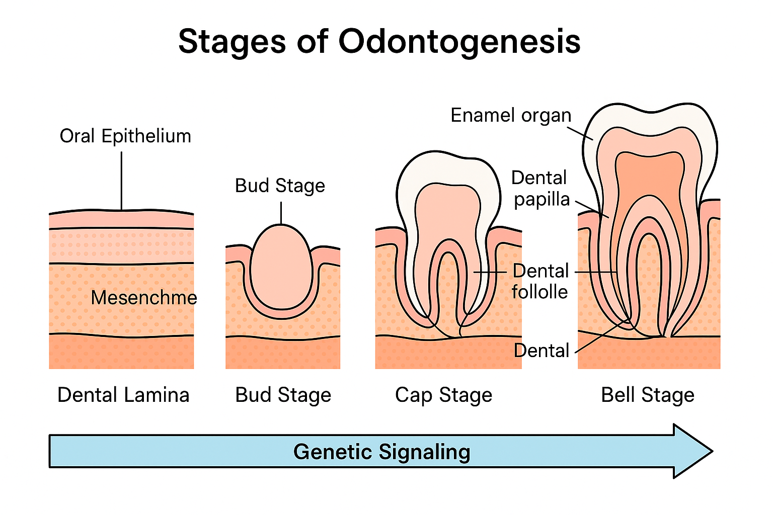
An Overview of Odontogenesis: The Principle of Tooth Formation
Odontogenesis is a complicated, multiphase process by which teeth grow out of embryonic cells into their complete form with the ability to chew. This biological wonder starts in the sixth week of the embryonic life and follows a path of five main stages namely the dental lamina, bud, cap, bell and maturation stages.
Each of the stages is broken down in detail here in this guide to odontogenesis.
The formation of teeth is dependent upon bidirectional communication between the oral epithelium (forming enamel) and the mesenchymal underlying the epithelium (forming dentin, pulp and cementum). These epithelial-mesenchymal interactions are strictly controlled by some gene families and molecular signal pathways, so that every tooth is formed in the proper shape, size, and location.
The Tooth Development Genetic Blueprint
The formation of teeth is not by chance but is preprogrammed. There are estimated to be over 300 genes that are involved in the process of odontogenesis, acting in separate, though overlapping, networks of signaling. Such genes may be classified by the developmental stages and molecular functions.
Key Gene Families Involved
Homeobox (HOX) Genes:
Homeobox genes are important in shaping the body and specifying where teeth are to grow. Namely, the formation of early tooth buds requires MSX1 and PAX9. The tooth agenesis is also commonly related to mutations in these genes in which one or more teeth do not develop.
Bone Morphogenetic Proteins (BMPs):
BMPs are signaling molecules which mediate cell-cell communication between epithelial and mesenchymal cells. They assist in governing cell proliferation and differentiation in order to bud-to-cap transition. BMP interference may lead to defective crowns and roots.
Fibroblast Growth Factors (FGFs):
FGFs particularly FGF8 and FGF10 also play a role in determining the shape and size of teeth. These molecules induce the proliferation and differentiation of mesenchymal cells, as well as odontoblast differentiation.
Sonic Hedgehog (SHH) Pathway:
SHH gene is the gene which regulates the growth of tooth germs and formation of enamel organs. This gene may undergo mutation and result in fused teeth or abnormal formulations of the enamel cover.
WNT Signaling Pathway:
WNT proteins influence several steps in the tooth morphogenesis. WNT10A and AXIN2 mutations have been associated with hypodontia and other developmental defects.
Phases of Genetic Control in Tooth Creation
Tooth formation may be separated into three large genetic control stages, which are initiation, morphogenesis, and differentiation.
Initiation Stage
At this initial step, FGF8, PAX9 and MSX1 start interaction between the oral epithelium and the mesenchymal cells. This will dictate where the tooth will be in future and the quantity.
Either a gene defect in MSX1 or PAX9 may result in non-syndromic tooth agenesis, usually of a second premolar or a third molar. These mutations disrupt the signaling cascade which induces development of the tooth buds.
Morphogenesis Stage
The tooth germ assumes a characteristic shape at the levels of the cap and the bell stage. SHH and BMP4 are genes that perfect cusp formation and general crown form. These genetic instructions cause the differentiation of enamel organ, dental papilla and dental follicle.
Mutations in SHH expression may result in either an abnormally-shaped denture (as in peg-shaped incisors) or even cell fusion of tooth germs, whereas mutations in BMP4 can lead to uneven thickness of the enamel.
Mineralization and Differentiation
The AMELX, ENAM, and DSPP genes control the mineralization of dentin and enamel after the crown form is set.
- A vital enamel matrix protein that regulates crystal growth is encoded by the amelogenin gene, or AMELX.
- The enamel rod’s hardness and organization are influenced by the enamelin gene, or ENAM.
- Dentin mineralization and tubule formation depend on the dentin sialophosphoprotein gene, or DSPP.
Amelogenesis imperfecta, or defective enamel formation, and dentinogenesis imperfecta, or abnormal dentin, are caused by mutations in these genes.
Dental Abnormalities and Genetic Mutations
Odontogenic gene mutations may influence the quantity and the architecture of teeth. Such genetic changes may be in the form of clinically identifiable disorders.
Amelogenesis Imperfecta (AI)
It is a hereditary disorder that is characterized by impaired enamel formation owing to mutation in AMELX, ENAM, or MMP20. The enamel becomes thin, soft or discoloured making teeth susceptible to wear and sensitivity.
- X-linked AI: This is caused by mutations in AMELX which result in generalized enamel hypoplasia.
- Autosomal-dominant AI: It is typically caused by mutations in ENAM, resulting in localized enamel defects.
Dentinogenesis Imperfecta (DI)
DI is an inherited condition that is caused by mutations in the DSPP gene, which is associated with discolored and translucent teeth and dentin defects. The enamel can be present but it breaks easily because of the defective underlying dentin.
Hypodontia and Tooth Agenesis
Missing teeth are known to be caused by loss- of-function mutations in MSX1, PAX9, WNT10A and AXIN2. The AXIN2 mutations are also interesting as they also correlate with a higher risk of colorectal cancer- indicating that odontogenesis genes can have an effect on the overall systemic health.
Supernumerary Teeth and Fusion Supernumerary
Teeth and Fusion Supernumerary teeth and fusion are features of the front teeth and molars that are absent in most mammals with a dental system like humans.
On the other hand, over activation of SHH or WNT signaling pathways can cause additional teeth (hyperdontia) or teeth fusion. These examples highlight the sensitivity at which genes need to be expressed during morphogenesis.
Odontogenesis Molecular Signaling Pathways
A number of molecular signaling pathways collaborate to help in the coordination of odontogenesis. The cross-talk they have provides appropriate differentiation and timing.
BMP and FGF Interactions
The relationship between BMP and FGF pathways is reciprocity between inhibition and stimulation. An illustration is that underdeveloped tooth germs that are spaced properly are regulated by the FGF8 expression that is inhibited by BMP4.
SHH and WNT Crosstalk
The pathways collaborate in the regulation of the patterning of the crown and enamel knot activity. The enamel knot is a signaling node that regulates the formation of the cusp. The impairment of both the pathways leads to abnormal crowns and cusp count.
Notch Signaling
The balance between the ameloblast and odontoblast differentiation is controlled by notch receptors and ligands (including JAG1). Mixed enamel-dentin anomalies may result because of notch mutations.
Regulation of Enamel and Dentin by Genetics.
Enamel Formation
Enamel matrix proteins, including amelogenin, enamelin and ameloblastin, are the key factors that regulate enamel formation which is also known as amelogenesis.
- Amelogenin (AMELX): Gives guidance to the structure of the hydroxyapatite crystal.
- Enamelin (ENAM): Supports the structure of the enamel rod.
- MMP20: Breaks down enamel proteins so that they can be mineralized.
Abnormalities in these genes cause hypo- or hypomineralized enamel commonly observed as white, yellow or brown discolouration.
Dentin Formation
Dentinogenesis is a differentiation process of the odontoblasts accompanied by the release of dentin matrix proteins:
- DSPP and its derivatives (DSP and DPP) are very important to mineralized dentin.
- Type I collagen that is coded by COL1A1 and COL1A2 gives structural strength.
Mutations in this case may result in weak dentin, large chamber of the pulp and bulbous crowns.
Morphology and Evolutionary Genetics of Teeth
The shape and arrangement of a tooth is also genetically predetermined. Minimal differences in cusp designs and root shapes are based on minor shifts in gene expression levels in the bell administration. An example to this is that EDA and SHH expression differences help to diversify molar cusps.
Evolutionarily, the reduction in tooth count and form in different species is linked to dietary changes – showing that minor genetic modifications can cause major morphological changes.
Genetic Testing and Individualized Dental Service
Genetic sequencing has provided new opportunities in the diagnosis of inherited dental diseases. The dentists are now able to detect particular mutations of genes that cause deformities, thus resulting in the more accurate treatment plan.
For example:
- Protective sealant and fluoride therapy might work with a patient with ENAM mutation.
- A PAX9 mutation child can be followed up at an early age due to lack of permanent teeth and provided with orthodontic instructions.
Gene therapy and regenerative dentistry could enable the repair of a defective enamel or even entire teeth with the aid of stem cell based bioengineering, based on the same genetic principles that govern natural odontogenesis.
Conclusion
Tooth development is genetic precision at its finest. Odontogenesis is the result of a complex interplay of genes and signaling pathways, which dictate the differentiation of uncommitted cells into the highly mineralized structures that contribute to our smiles.
A mutation in a single gene, such as AMELX, PAX9 or DSPP, may result in significant alterations in the quality or quantity of enamel and dentin, or in the number of teeth. Reading out these genetic blueprints, dentistry today is transitioning from mechanical restoration to biologically informed prevention and regeneration.
Understanding tooth’s genetic basis not only helps us better appreciate the wonder of human development, it paves the way for personalized dental care in the future, where every treatment plan matches each patient’s individual genetic signature.