Fluorescence microscopy is a pivotal technique in modern biological and medical research, enabling detailed visualization and analysis of cellular structures and processes. This article delves into its fundamental principles, components, types, operating procedures, applications, advantages, limitations, precautions, and notable examples of fluorescence microscopes.
Whether you’re a budding scientist, a curious student, or a professional researcher, FAQs on microscopelog offer comprehensive information on the various types of microscopes, their uses, and the incredible discoveries they enable. From basic principles to advanced techniques, you’ll find everything you need to expand your knowledge and appreciation of this essential scientific tool. Explore today and unlock the secrets hidden in the smallest details!
Basic Principle
At its core, fluorescence microscopy leverages the fluorescence phenomenon. Fluorophores within biological samples absorb light at a specific excitation wavelength, subsequently emitting light at a longer wavelength known as the emission wavelength. This emitted light is then captured to form high-resolution images, highlighting specific structures labeled with fluorophores against a dark background.
Components of a Fluorescence Microscope
Key components include:
- Light Source: The light source in fluorescence microscopy is critical for providing the excitation light necessary to activate fluorophores within the sample. Common sources include mercury lamps and lasers, which emit intense light at specific wavelengths suitable for exciting fluorophores.
- Excitation Filters: These filters are positioned in the light path to selectively transmit the excitation wavelengths of light emitted by the source. By filtering out unwanted wavelengths, excitation filters ensure that only the light necessary to excite the fluorophores reaches the sample.
- Dichroic Mirror (Beam Splitter): Positioned at a 45-degree angle to the light path, the dichroic mirror reflects the excitation light towards the sample while allowing emitted fluorescence to pass through unimpeded. This optical component plays a crucial role in separating the excitation and emission wavelengths of light.
- Objective Lens: The objective lens collects the emitted fluorescence from the sample and focuses it onto the detector. This lens is designed to gather as much light as possible while maintaining high resolution, enabling detailed imaging of the fluorescently labeled structures within the sample.
- Emission Filters: Similar to excitation filters, emission filters are used to selectively transmit only the emitted fluorescence wavelengths. By blocking out the excitation light and allowing only the emitted light to pass through, emission filters help to enhance contrast and improve the quality of the final image.
- Detectors: Detectors in fluorescence microscopy are crucial for converting the emitted fluorescence light into measurable signals. Common types include CCD cameras and photomultiplier tubes (PMTs), which capture and amplify the weak fluorescence signals, respectively, ensuring high sensitivity and resolution in imaging.
Detectors in Fluorescence Microscopy
Detectors in fluorescence microscopy are crucial for converting emitted fluorescence light into measurable signals. Common types include:
- CCD Cameras: Charge-Coupled Devices (CCDs) are widely used in fluorescence microscopy due to their ability to capture images with high spatial resolution and sensitivity. These detectors convert photons of emitted fluorescence into electronic signals, which are then processed to create digital images. CCD cameras are suitable for various fluorescence imaging applications, offering versatility and reliability in research and clinical settings.
- Photomultiplier Tubes (PMTs): Photomultiplier Tubes are highly sensitive detectors used to amplify weak fluorescence signals in fluorescence microscopy. PMTs convert individual photons into electrical signals through a series of electron multiplication stages, resulting in significantly enhanced signal-to-noise ratios and rapid response times. This makes PMTs particularly suitable for applications requiring detection of low-intensity fluorescence signals or dynamic processes in live cell imaging.
Wavelengths Used in Fluorescence Microscopy
In fluorescence microscopy, excitation wavelengths typically range from ultraviolet (UV) to near-infrared (NIR), depending on the fluorophore’s absorption properties. Emission wavelengths are longer than excitation wavelengths and correspond to the specific fluorescence emitted by the fluorophore.
Light Path of a Fluorescence Microscope
The light path in a fluorescence microscope involves several critical steps:
- Excitation: Light from the source passes through the excitation filter, which selects a specific wavelength to excite fluorophores in the sample.
- Dichroic Mirror: Reflects the excitation light towards the sample, while allowing emitted fluorescence to pass through.
- Objective Lens: Collects and focuses emitted fluorescence from the sample.
- Emission: Fluorescence passes through an emission filter, blocking excitation light and allowing only emitted fluorescence to reach the detector for image formation.
Types of Fluorescence Microscopes
Different types cater to various imaging needs:
- Wide-field Epifluorescence Microscope: Simultaneously illuminates the entire sample, ideal for rapid imaging of thin specimens.
- Confocal Microscope: Utilizes a pinhole to eliminate out-of-focus light, enabling high-resolution optical sectioning and 3D imaging.
- Multiphoton Microscope: Uses longer-wavelength light for deeper tissue penetration and reduced photodamage.
- Total Internal Reflection Fluorescence Microscope (TIRF): Selectively excites fluorophores near the specimen surface, perfect for studying cell membrane dynamics.
Applications of Fluorescence Microscopy
Applications span various fields:
- Cell Biology: Visualizing cellular structures, organelles, and dynamic processes.
- Molecular Biology: Studying protein localization, gene expression, and molecular interactions.
- Neuroscience: Imaging neuronal structures and activities.
- Medical Diagnostics: Detecting biomarkers for disease diagnosis and treatment monitoring.
Advantages of Fluorescence Microscopy
Fluorescence microscopy offers several advantages:
- High Sensitivity: Detects single molecules with high sensitivity.
- Specificity: Targets specific structures or molecules within complex samples.
- Multicolor Imaging: Simultaneously visualizes multiple fluorescent labels.
- Live Cell Imaging: Observes dynamic processes in real-time without disrupting cellular activities.
Limitations and Precautions
However, fluorescence microscopy has limitations:
- Photobleaching: Fluorophore degradation due to prolonged exposure to excitation light.
- Phototoxicity: Potential cell damage from high-intensity light.
- Background Fluorescence: Autofluorescence from samples can interfere with specific signal detection.
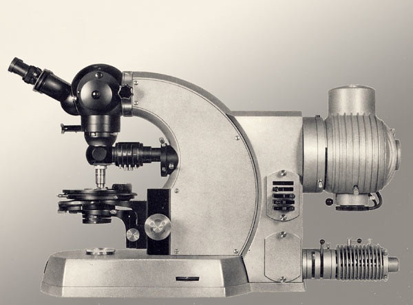
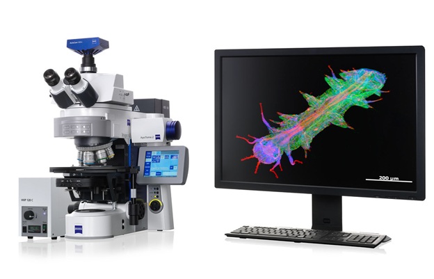
Examples of Fluorescence Microscopes
Advanced models include:
- Optical Microscope Dragonfly 600 (Andor Technology PLC): Known for high-speed imaging.
- Optical Microscope MSC- B107T (Infitek): Versatile for research and clinical settings.
- Fluorescence Microscope DM6 FS (Leica): Automation and high-resolution imaging capabilities.
- Optical Microscope MiOF-500N (Microptik BV): Compact and efficient for diverse applications.
Conclusion
Fluorescence microscopy remains indispensable for advancing scientific understanding across disciplines. By mastering its principles, researchers unlock powerful tools for exploring cellular intricacies and pushing the boundaries of biomedical knowledge. Continued innovation in technology and methodology ensures fluorescence microscopy’s vital role in driving discoveries and improving healthcare outcomes.